since 09. July 2013
C O N T E N T S 3.2013
COMMENTS ON THE ARTICLES
ORIGINAL ARTICLES
Jeetun Oomar, Jeewon Rajesh
A survey of nail infection and awareness among non-diabetic patients in Mauritius
Our Dermatol Online 2013; 4(3): 265-271 DOI: 10.7241/ourd.20133.65
[abstract-English] , [article in English], [PDF], [HTML]
Nermina Ovcina-Kurtovic, Emina Kasumagic-Halilovic
Prevalence of nail abnormalities in patients with psoriasis
Our Dermatol Online 2013; 4(3): 272-274 DOI: 10.7241/ourd.20133.66
[abstract-English] , [article in English], [PDF], [HTML]
Ana Maria Abreu Velez, Maria Mercedes Yepes Naranjo, Isabel Cristina Avila, Martha Luz Londoño, Paul B. Googe Jr., Jorge Enrique Velásquez Velez, Ivan Dario Velez, Yulieth Alexandra Upegui, Alejandra Jimenez- Echavarria, Natalia Regina Mesa-Herrera, Hong Yi, Juliana Calle-Isaza, Michael S. Howard
Tissue inhibitor of metalloproteinase 1, matrix metalloproteinase 9, αlpha-1 antitrypsin, metallothionein and urokinase type plasminogen activator receptor in skin biopsies from patients affected by autoimmune blistering diseases
Our Dermatol Online 2013; 4(3): 275-280 DOI: 10.7241/ourd.20133.67
[abstract-English] , [article in English], [PDF], [HTML]
Toraub Kawshar, Jeewon Rajesh
Sociodemographic factors and their association to prevalence of skin diseases among adolescents
Our Dermatol Online 2013; 4(3): 281-286 DOI: 10.7241/ourd.20133.68
[abstract-English] , [article in English], [PDF], [HTML]
Maryana Kovalchuk, Mariia Shkilna, Mykhailo Andreychyn, Natalia Vasylieva, Vasyl Demyanenko
Optimization of lambliasis microscopic diagnostics by the method of polarized fluorescence for patients with rosacea and urticaria
Our Dermatol Online 2013; 4(3): 287-289 DOI: 10.7241/ourd.20133.69
[abstract-English] , [article in English], [PDF], [HTML]
Neerja Puri, Asha Puri
A study on topical calcium dobesilate for the treatment of limited plaque psoriasis
Our Dermatol Online 2013; 4(3): 290-293 DOI: 10.7241/ourd.20133.70
[abstract-English] , [article in English], [PDF], [HTML]
Deeptara Pathak Thapa, Anil Kumar Jha
Clinico – histopathological correlation in leprosy: a tertiary care hospital based study
Our Dermatol Online 2013; 4(3): 294-296 DOI: 10.7241/ourd.20133.71
[abstract-English] , [article in English], [PDF], [HTML]
Kotowaroo Goonmatee, Jeewon Rajesh
What factors contribute to a higher frequency of skin infections among adults in Mauritius?
Our Dermatol Online 2013; 4(3): 297-302 DOI: 10.7241/ourd.20133.72
[abstract-English] , [article in English], [PDF], [HTML]
Neerja Puri, Asha Puri
A study on lichen planus in children
Our Dermatol Online 2013; 4(3): 303-305 DOI: 10.7241/ourd.20133.73
[abstract-English] , [article in English], [PDF], [HTML]
Kallappa C. Herkal, Suma Patil, Hosahalli Rjaiah Yogeesh, Raghu Muddigere Thimmappa, Lalitha Cholachaguddar
Study of therapeutic comparison of tacrolimus 0.1% and minoxidil 2% in alopecia areata
Our Dermatol Online 2013; 4(3): 306-310 DOI: 10.7241/ourd.20133.74
[abstract-English] , [article in English], [PDF], [HTML]
Neerja Puri, Asha Puri
A clinical and histopathological study of cicatricial alopecia
Our Dermatol Online 2013; 4(3): 311-315 DOI: 10.7241/ourd.20133.75
[abstract-English] , [article in English], [PDF], [HTML]
CASE REPORTS
Neerja Puri
Recalcitrant widespread alopecia areata in a child treated successfully with oral methylprednisolone pulse therapy
Our Dermatol Online 2013; 4(3): 316-318 DOI: 10.7241/ourd.20133.76
[abstract-English] , [article in English], [PDF], [HTML]
Erick Francisco Sanchez Jimenez
Erythema Elevatum Diutinum as most probable diagnosis: a case report
Our Dermatol Online 2013; 4(3): 319-321 DOI: 10.7241/ourd.20133.77
[abstract-English] , [article in English], [PDF], [HTML]
Verónica Uraga, Andrea Lubkov, Annette Morán, Juan Carlos Garcés, Enrique Uraga
Granular Parakeratosis: Report of 2 Ecuadorian cases and Review of the Literature
Our Dermatol Online 2013; 4(3): 322-324 DOI: 10.7241/ourd.20133.78
[abstract-Engli sh] , [article in English], [PDF], [HTML]
Ashutosh Talwar, Neerja Puri
Kimura’s disease – a rare entity
Our Dermatol Online 2013; 4(3): 325-327 DOI: 10.7241/ourd.20133.79
[abstract-English] , [article in English], [PDF], [HTML]
Vikram K. Mahajan, Pushpinder S. Chauhan, Karaninder S. Mehta, Vikas Sharma
Our Dermatol Online 2013; 4(3): 328-329 DOI: 10.7241/ourd.20133.80
[abstract-English] , [article in English], [PDF], [HTML]
Anca Chiriac, Liliana Foia, Anca E Chiriac, Cristina Birsan, Caius Solovan, Piotr Brzezinski
Lichen striatus – case reports
Our Dermatol Online 2013; 4(3): 330-332 DOI: 10.7241/ourd.20133.81
[abstract-English] , [article in English], [PDF], [HTML]
Chebbi Wafa, Ajili Faida, Boussetta Najeh, Abderrezak Fatma, Othmani Salah, Sfar Mohamed Habib
Erythema nodosum revealing acute myeloid leukemia
Our Dermatol Online 2013; 4(3): 333-334 DOI: 10.7241/ourd.20133.82
[abstract-English] , [article in English], [PDF], [HTML]
Geetha Krishnanand, Vidya Monappa, Anuradha C.K. Rao
Primary cutaneous NK/T cell lymphoma-nasal type with cutaneous aspergillosis. A case report and literature review
Our Dermatol Online 2013; 4(3): 335-338 DOI: 10.7241/ourd.20133.83
[abstract-English] , [article in English], [PDF], [HTML]
Comment by: Ass. Prof. Małgorzata Sokołowska-Wojdyło
Our Dermatol Online 2013; 4(3): 339-340 DOI: 10.7241/ourd.20133.83.1
[article in English], [PDF], [HTML]
Taeko Nakamura-Wakatsuki, Toshiyuki Yamamoto
Periorbital necrobiotic xanthogranuloma without paraproteinemia
Our Dermatol Online 2013; 4(3): 341-343 DOI: 10.7241/ourd.20133.84
[abstract-English] , [article in English], [PDF], [HTML]
Manuel Valdebran, Rogelio Isao Salinas, Nelly Ramirez, Alba Rodriguez, Leyla Guzman, Silvia Marte, Max Suazo, Esmirna Rosado
Fixed drug eruption of the eyelids. A dermoscopic evaluation
Our Dermatol Online 2013; 4(3): 344-346 DOI: 10.7241/ourd.20133.85
[abstract-English] , [article in English], [PDF], [HTML]
Tomoko Oishi, Yuka Hanami, Yasunobu Kato, Mikio Otsuka, Toshiyuki Yamamoto
Staphylococcal scalded skin syndrome mimicking toxic epidermal necrolysis in a healthy adult
Our Dermatol Online 2013; 4(3): 347-348 DOI: 10.7241/ourd.20133.86
[abstract-English] , [article in English], [PDF], [HTML]
Sonia Bhatt, Nalini Bhaskaranand, Kashyap Udupa, Meenu Joon
Progressive varicella syndrome with Varicella gangrenosa in an immune-competent infant
Our Dermatol Online 2013; 4(3): 349-350 DOI: 10.7241/ourd.20133.87
[abstract-English] , [article in English], [PDF], [HTML]
Swati Sharma, Manna Valiathan, Sushama Belurkar, Raviraj Acharya, Swati Aggarwal
Cytophagic histiocytic panniculitis associated with HBe hemoglobinopathy in a patient with hemophagocytic syndrome
Our Dermatol Online 2013; 4(3): 351-353 DOI: 10.7241/ourd.20133.88
[abstract-English] , [article in English], [PDF], [HTML]
Comment by: Prof. Hubert Daisley
Our Dermatol Online 2013; 4(3): 354-355 DOI: 10.7241/ourd.20133.88.1
[article in English], [PDF], [HTML]
Kanthilatha Pai, Sathish Pai
Localised involutional lipoatrophy: a case report
Our Dermatol Online 2013; 4(3): 356-357 DOI: 10.7241/ourd.20133.89
[abstract-English] , [article in English], [PDF], [HTML]
Lourdes Rodríguez, Beatriz Di Martino Ortiz, Romina Contreras, Mirtha Rodriguez Masi, Oilda Knopfelmacher, Lourdes Bolla de Lezcano
Calcinosis cutis metastásica: Calcifilaxis (arteriolopatía urémica calcificada). A propósito de un caso
Metastatic calcinosis cutis: calciphylaxis (calcified uremic arteriolopathy). A case report
Our Dermatol Online 2013; 4(3): 358-360 DOI: 10.7241/ourd.20133.90
[abstract-Spanish, English] , [article in Spanish], [PDF], [HTML]
Fatou Barro/Traoré, Myriam Sanwidi, Fousséni Dao, Nina Korsaga/Somé, Pascal Niamba, Adama Traoré, Ludovic Kam, Gilles Lemasson
Histoplasmose africaine disséminée chez un enfant immunocompétent au Burkina Faso: un cas
Disseminated African Histoplasmosis in an immunocompetent child in Burkina Faso: one case
Our Dermatol Online 2013; 4(3): 361-368 DOI: 10.7241/ourd.20133.91
[abstract-French, English] , [article in French], [PDF], [HTML]
REVIEW ARTICLES
Iffat Hassan, Konchok Dorjay, Abdul Sami, Parvaiz Anwar
Sunscreens and Antioxidants as Photo-protective Measures: An update
Our Dermatol Online 2013; 4(3): 369-374 DOI: 10.7241/ourd.20133.92
[abstract-English] , [article in English], [PDF], [HTML]
Yogeesh Hosahalli Rajaiah, Kallappa C. Herkal
Ploymorphous Light Eruption – a review
Our Dermatol Online 2013; 4(3): 375-379 DOI: 10.7241/ourd.20133.93
[abstract-English] , [article in English], [PDF], [HTML]
CLINICAL IMAGES
Patricia Chang
Molluscum Contagiosum of scalp in a child
Our Dermatol Online 2013; 4(3): 380-381 DOI: 10.7241/ourd.20133.94
[article in English], [PDF], [HTML]
LETTER TO THE EDITOR
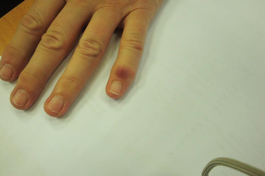
Sunil Kumar, Sanjay Diwan, Milind Dekate, Sonam Goyal
Mees’ line following chemotherapy
Our Dermatol Online 2013; 4(3): 382 DOI: 10.7241/ourd.20133.95
[article in English], [PDF], [HTML]
HISTORICAL ARTICLES
Khalid Al Aboud, Ahmad Al Aboud
Eponyms in the dermatology literature linked to the histiocytic disorders
Our Dermatol Online 2013; 4(3): 383- 384 DOI: 10.7241/ourd.20133.96
[article in English], [PDF], [HTML]
Khalid Al Aboud, Daifullah Al Aboud
Eponyms in the literature of cutaneous lymphomas
Our Dermatol Online 2013; 4(3): 385-388 DOI: 10.7241/ourd.20133.97
[article in English], [PDF], [HTML]
Khalid Al Aboud, Ahmad Al Aboud
Eponyms in the dermatology literature linked to the skin and soft tissue tumors
Our Dermatol Online 2013; 4(3): 389-391 DOI: 10.7241/ourd.20133.98
[article in English], [PDF], [HTML]
Khalid Al Aboud, Ahmad Al Aboud
Eponyms in the dermatology literature linked to the vascular tumors
Our Dermatol Online 2013; 4(3): 392-394 DOI: 10.7241/ourd.20133.99
[article in English], [PDF], [HTML]
Khalid Al Aboud, Daifullah Al Aboud
Eponyms in the dermatolopathology literature linked to the neural tissues
Our Dermatol Online 2013; 4(3): 395-398 DOI: 10.7241/ourd.20133.100
[article in English], [PDF], [HTML]
Piotr Brzezinski, Anca Chiriac, Cecil Munsey, Ahmad Thabit Sinjab
Dermatology Eponyms – sign –Lexicon (J)
Our Dermatol Online 2013; 4(3): 399-402 DOI: 10.7241/ourd.20133.101
[abstract-English] , [article in English], [PDF], [HTML]
COMMENTS ON THE ARTICLES
PREVALENCE OF NAIL ABNORMALITIES IN PATIENTS WITH PSORIASIS
Dr. Rajesh Jeewon Ph.D. (Mauritius)
This is an interesting article that contributes a better understanding of nail abnormalities about patients with psoriasis. Obviously there are multifactorial factors that can lead to an increased incidence of onychomycosis among this selected target population. Recent studies have demonstrated that there is an increase in the prevalence of Onychomycosis among psoriatic patients. The authors also attempted to correlate different factors to nail abnormalities. Males appear to be more vulnerable than females and this has been shown in other studies as well. The main fungi associated with nail abnormalities have also been shown to be Candida albicans and Trichophyton Mentagrophytes and similar results herein support previous studies.
Prof. Uwe Wollina (Germany)
Nail changes in psoriasis have been described a long time ago. Their treatment has been frustrating. So interest in nail changes has dropped down.
Only recently a new interest in psoriatic nails has been fueled by progress in molecular pathology and inflammation and the availability of targeted drugs. Although nail changes can be observed in both psoriasis of skin and psoriatic arthritis, a lot of new findings have been gathered from the last. In particular the interrelationship of peripheral arthritis, daktylitis and enthesitis to nail changes has been discussed intensively. The question of hen and egg has been largely been resolved. It seem clear now, that the affection of distal interphalangeal joints and entheses provides the background of a higher incidence of nail changes as long as the arthritis is active. Other types of psoriatic arthritis, i.e. spine involvement or affection of large joints, do not demonstrate such a close relationship to the nails. N. Ovcina- Kurtovic and E. Kasumagic-Halilovic investigated the point prevalence of nail changes in patients with psoriasis and psoriatic arthritis in Bosnia and Hercegovina. Two-third of their patients presented with nail changes, pitting and discoloration as the most common clinical symptoms.
In general none of the different types of psoriatic disorders has a particular prevalence to nail changes (except active peripheral arthritis and enthesitis) [1].
References:
1. Wollina U, Barta U, Uhlemann C, Oelzner P, Hein G: Nail changes in rheumatoid arthritis. Hautarzt. 1999;50:549-55.
Prof. Iffat Hassan (India)
The authors need to be complimented on dwelling on a very important clinical problem that one encounters in day to day clinical practice. However, it would have been interesting if simple clinical tools like Nail Psoriasis Severity Index (NAPSI) or the modified NAPSI which are numeric, reproducible, objective, simple tools for evaluation of nail psoriasis were also put into practice while examining the nails in psoriasis patients.
A STUDY ON TOPICAL CALCIUM DOBESILATE FOR THE TREATMENT OF LIMITED PLAQUE PSORIASIS
Ass. Prof. Belliappa Pemmanda Raju (India)
I congratulate the authors for doing a commendable work. The article throws light on the safety and efficacy of calcium dobesilate in limited plaque psoriasis. Results look promising with the least side effect profile compared to steroids and other topicals. However, longer follow-up is required to see for the disease free remission period it offers. Further studies are required on a larger population.
The drug used was potassium or calcium dobesilate? few areas it has been mentioned as potassium!
A STUDY ON LICHEN PLANUS IN CHILDREN
Ass Prof. Nagat Sobhy Mohamad (Egypt)
This study direct our attention to the occurance of lichen planus in children and how to treat.
RECALCITRANT WIDESPREAD ALOPECIA AREATA IN A CHILD TREATED SUCCESSFULLY WITH ORAL METHYLPREDNISOLONE PULSE THERAPY
Prof. Sunderamoorthy Srinivasan (India)
Methylprednisolone was not based on the weight of the patient. The two pictures have been taken on the same day, post-treatment picture is not available. Please clarify.
ERYTHEMA ELEVATUM DIUTINUM AS MOST PROBABLE DIAGNOSIS: A CASE REPORT
Anca Chiriac, MD PhD and Prof. Caius Solovan (Romania)
It is a well documented presentation, up to date, very clear and didactic paper, a concise case,that tries to make more light in difficult and rare dermatological problems.
It was also an opportunity for us to re-evaluate some aspects, to recall some cases from own experience.
We would like just to emphasize some important points in daily practice, most of them already discussed in the paper.
Erythema elevatum diutinum is indeed a rarely reported disease, especially in children, most cases being presented to adults.
It has been described in the literature under different terms: Erythema annulare centrifugum, Erythema figuratum perstans, Erythema gyratum perstans, Erythema, annulare centrifugum, Erythema, gyratum perstans.
The hallmark of the disease is the striking clinical aspect of the lesions (one can be remembered for always in typical cases): plum colored nodules and plaques, associated with minimal symptoms, distributed symmetrically over the extensor surfaces of extremities, most often hands and feet, but also knees, elbows, buttocks, even face and ears.
The evolution of the lesions is quite characteristic also: sometimes with restitution ad integrum but more often to atrophic or fibrotic scars and hyperpigmentation.
Although the clinical picture is quite helpful for diagnosis the definitive conclusion is made after histopathological report that emphases the aspect of leukocytoclastic vasculitis and the dermal neutrophilic infiltrate.
Sometimes the clinico-pathological report must eliminate others morbidities: sarcoidosis, discoid erythematous lupus, erythema multiforme minor type, leprosy, Sweet syndrome or other forms of small vessel vasculitis, Kaposi sarcoma, especially in HIV positive patients with Erythema elevatum diutinum lesions on the lower extremities (Kaposi sarcoma presents lesion in the oral cavity unusual to classic form with lesion on the lower extremity like in Erythema elevatum diutinum with HIVpositive patients).
Taken into consideration the variety of the diseases reported being associated with Erythema elevatum diutinum:
· autoimmune diseases (rheumatoid arthritis, celiac and inflammatory bowl disease, diabetes mellitus),
· infections :syphilis, streptococcus, hepatitis, HIV infection),
· malignancies :multiple myeloma, B-cell lymphoma, breast carcinoma, myelodysplasia,
· monoclonal gammopathies, hypergammaglobulinemia,
· pyoderma gangrenosum,
· polychondritis. systemic lupus erythematosus; Sjögren’s syndrome, juvenile idiopathic arthritis
the paraclinic examinations must be carefully selected and a thorough interpretation must be of value.
For example: some rheumatologic disorders (Behçet’s disease, cryoglobulinemic vasculitis, Henoch-Schönlein purpura, hypersensitivity vasculitis) may exhibit leukocitoklastic vasculitis and the diagnosis can be wrong.
Immunoelectrophoresis testing must be recommended to all cases because of IgA monoclonal
gammopathy frequently reported in cases of Erythema elevatum diutinum.
Glucose 6-phosphate dehydrogenase is compulsory to have it done for Dapsone treatment still considered nowadays the most successful therapy in Erythema elevatum diutinum.
A large battery test is needed to elucidate the co morbidities or associations:creatine phosphokinase; lactic dehydrogenase; VDRL; ASO; thyroid hormones; serology for HIV and hepatitis; rheumatoid factor; Waaler-Rose test; antinuclear antibodies; autoantibodies (anti-Ro, anti-La, anti-DNA and anti-RNP antibodies); cryoglobulins and cryoagglutinins; chest radiography; and the list can continue.
Systemic corticosteroids are not effective even in early stages; Dapsone does not resolve all the cases (one can see the long list of other proposed and tried therapeutic options)
Do not forget the antiretroviral treatment in HIV associated Erythema elevatum diutinum.
We congratulate the authors for the diagnosis and presentation.
Dr Marko Vok (Slovenia)
This case report is very interesting, but it is extremely difficult to say what is really this skin eruption. Erythema elevatum diutinum (EED) is a chronic condition. Here we have a presentation of a very acute condition localized on the palms. EED is usually distributed over extensor surfaces, although cases on palms and soles have also been reported.
Histologic examination in EED shows a constellation of nonspecific signs. Acute lesions of EED demonstrate leukocytoclastic dense dermal neutrophilic infiltrate, parietal fibrinoid vascular necrosis and collagen fiber necrosis. Eosinophils may also be presented in the upper and mid-dermis. Depending on the degree of oedema and infiltration into the dermis, unaffected collagen may be present just under the epidermis [1]. In this case report there are some elements of an early EED, but far from been enough for the diagnosis of EED. And there is a confusing epidermal pustula. EED is a chronic cutaneous vasculitis occurring in association with a variety of conditions including autoimmunity, infectious diseases and hematological abnormalities. The role of associated diseases is controversial, and the exact pathogenesis of EED is unknown. The association of EED with monoclonal or polyclonal IgA gammopathies seems to be important. The determination of antineutrophil cytoplasmic antibodies (ANCAs) could be helpful in the diagnosis of EED [2]. So, it would be advisable to perform some additional tests like: protein electrophoresis, indirect and direct immunofluorescence.
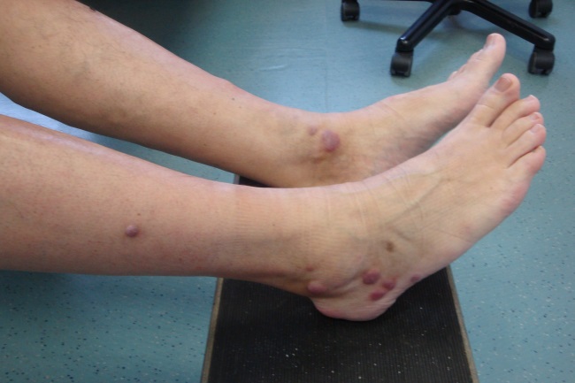
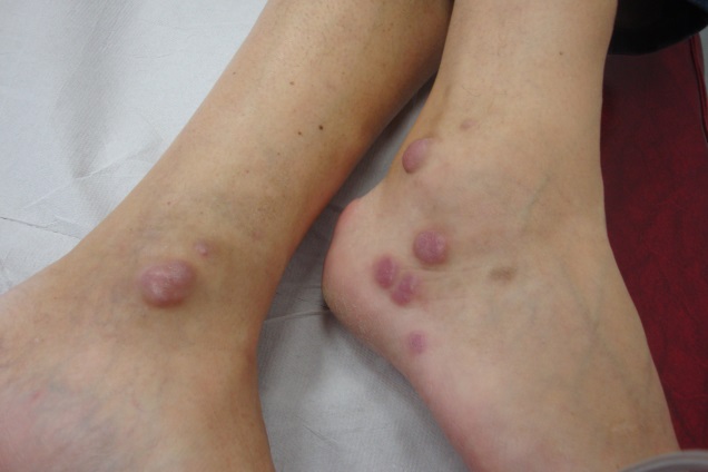
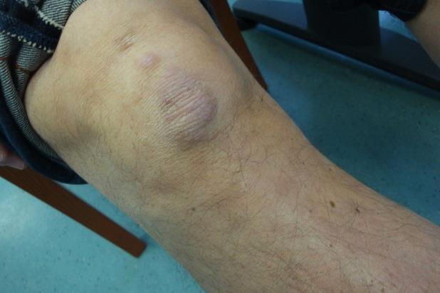
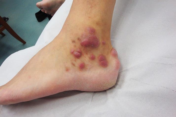
Diutinum in latin means lasting a long time. The expression is suitable to designate not only the disease, but also the time required for the diagnosis of EED. At the end of this comment I would like to add some images of my only 49-year old male patient with EED.
|
Figure 1. Plaques on the ankles
|
Figure 1. Plaques on the ankles
|
|
Figure 1. Plaques on the knee
|
Figure 1. Plaques on the ankle
|
|
Figure 1. A small plaque on the little finger
|
References
1. Barham K, Jorizzo J, Grattan B, Cox N: Vasculitis and neutrophilic vascular reations. In: Burns T, Breathnach S, Cox N, Griffiths C, eds. Rook’s Textbook of Dermatology. 7th ed. Blackwell Science; 2004.
2. Ayoub N, Charuel J-L, Diemert M-C, Barete S, Andre´ M, Fermand J-P, et al: Antineutrophil cytoplasmic antibodies of IgA class in neutrophilic dermatoses with emphasis on erythema elevatum diutinum. Arch Dermatol. 2004;140:931-6.
GRANULAR PARAKERATOSIS: REPORT OF 2 ECUADORIAN CASES AND REVIEW OF THE LITERATURE
Viktoryia Kazlouskaya, MD, PhD (Belarus)
Granular parakeratosis is a relatively rare and probably often misdiagnosed/underdiagnosed condition. It was described only a decade ago and we are still not aware of its spectrum of its clinical presentations and treatment modalities. The article gives us to opportunity to widen our horizons in understanding this condition, it presents also a case of granular parakeratosis in a child that is even more rare and unique.
PRIMARY CUTANEOUS NK/T CELL LYMPHOMA-NASAL TYPE WITH CUTANEOUS ASPERGILLOSIS. A CASE REPORT AND LITERATURE REVIEW
Drs. Luz Calderón, Rosa María Ponce and Alexandro Bonifaz (Mexico)
The article by Krishanand, Monappa and Rao presents a very interesting case report, showing the development of a primary cutaneous aspergillosis over one of the rarest types of T cell lymphoma.
The extranodal NK T cell lymphoma (NKTCL) refers to a group of clonal proliferations of cytotoxic lymphocytes of natural killer or T cell types, arising mainly in the nasal cavity. Nasal type extranodal NKTCL is characterized by the presence of malignant cells that are usually CD2 and CD56 positive (NK phenotype), with cytoplasmic, but not surface, CD3 positivity; which contain cytotoxic proteins such as T-cell intracellular antigen 1, perforin and granzyme B. Rarely, extranodal NKTCL may begin in other extranodal sites such as the skin, gastrointestinal tract, testis or lungs; being even more aggressive and with worst clinical outcome. The 5-year survival rate is less than 50%, with a median overall survival of 7.8 months, corresponding to the poorest survival among all T cell lymphomas [1,2].
Lymphoma and leukemia patients are at more risk for fungal infections. Opportunistic fungal infections are widely distributed, and frequent in the developing countries, the most common are due to Aspergillus spp. [3], related generally to neutropenia. It is possible to find primary cutaneous fungal infections and cutaneous manifestations of fungemia. Primary cutaneous aspergillosis is rare, both genders are equally affected. It starts as a result of the injuries caused by catheters, probes, needles and adhesive bandages. Initially, uncharacteristic erythematous papules appear and later become hemorrhagic plaques with necrotic areas. In this specific case the clinical suspicion is of special importance because the lymphoma itself manifests as ulcerating lesions, red plaques or nodules commonly ulcerated. Therefore, the patients require meticulous cleansing and aggressive antifungal therapy.
When it comes to immunosuppressed patients, dermatologists should always be aware of other opportunistic fungal infections such as mucormycosis, phaehyphomycosis and hyalohyphomycosis. Mucorales are worldwide distributed, by and large prefer humid environment. The most important risk factors are ketoacidosis, neutropenia, lymphoma, leukemia, malnutrition, burns, and use of deferoxamine. The rhinocerebral form is more frequent, while primary cutaneous mucormycosis are rare. Likewise starts after dermal injuries as an erythematous lesion that rapidly becomes necrotic with putrid malodorous discharge. Fusarium spp. infection develops mostly trough inhalation, but primary cutaneous forms are possible following direct inoculation, presenting as painful necrotic areas and ulcers [3].
Clinical diagnosis is difficult, and complimentary tests should always been used to determine the specific fungi and treatment, especially for the implications that come with the possible spread of a primary cutaneous fungal infection.
References
1. Schmitt C, Sako N, Bagot M, Huang Y, Gaulard P, Bensussan A: Extranodal NK/T-cell lymphoma: Toward the identification of clinical molecular targets. J Biomed Biotechnol. 2011;2011:190871.
2. Au WY, Weisenburger DD, Intragumtornchai T, Nakamura S, Kim WS, Sng I, et al. Clinical differences between nasal ans extranasal natural killer/T-cell lymphoma: a study of 136 cases from the International Periferal T-Cell Lymphoma Project. Blood. 2009;113:3931-7.
3. Perusquía-Ortiz AM, Vázquez-González D, Bonifaz A: Opportunistic filamentous mycoses: aspergillosis, mucormycosis, phaeohyphomycosis and hyalohyphomycosis. J Dtsch Dermatol Ges. 2012;10:611-21.
Dr. Mohamed Wael Daboul (Syrian Arab Republic)
A minor population of ‘lymphocytic’ cells which do not carry markers of either T or B cells are known as ‘non-T, non-B’ cells or ‘third population’ cells. The sequence of differentiation of these cells is not known. The majority of ‘null’ cells appear as large granular lymphocytes in the peripheral blood. This population of cells contains the majority of natural killer (NK) cells, which were originally described on a functional basis according to their capability of killing certain tumor cells of hematopoietic origin in the absence of prior stimulation or sensitization. Natural Killer cells (NK) are large cells with pale blue cytoplasm and a high cytoplasmic to nuclear ratio. These cells constitute 2 to 6% of the peripheral white cells and approximately 10 to 15% of the peripheral blood lymphocytes. Natural killer (NK) cells are antibody-independent cellular cytotoxic cells (AIDCC), and do not require major histocompatibility complex (MHC) recognition. NK cells are a heterogeneous population with respect to phenotype and target specificity. Although the majority of the CD56 + NK cells are CD3 -, small numbers of CD45 +/CD3 + cells have been detected and large granular lymphocyte (LGL) leukemias with the same phenotype have been reported. NK cells probably include cells of mainly T-cell (CD81), (CD161) and myeloid-cell lineages. Their proliferation is stimulated by interleukin-2, interleukin-4, interleukin-12, interleukin-13, and g-interferon.
The sequence of events in T cells development appears to be initial expression of nuclear terminal deoxynucleotidyl transferase (TdT) and the surface antigen CD7 followed by CD2. Mature T cells carry a marker (antigen CD2), which binds sheep erythrocytes. Adherence of thymocytes to epithelial cells is mediated by CD2. CD2 and CD7 expression on the cell surface may precede arrival in the thymus. Those early T cells are Potentially, capable to differentiate to other lineages such as natural killer (NK) cells. CD3 antigen are expressed on the surface later, although intracytoplasmic CD3 is one of earliest markers. Because of a common ontogeny, NK cells express T cell antigens, including CD2, CD7 and CD8. They are negative for surface CD3, but express cytoplasmic CD3 epsilon (e) chain. NK cells also express “NK-lineage associated” markers, including CD16, CD56 and CD57. Of these antigens, CD56 is generally regarded as an NK cell marker, although it can also be expressed on NK-like T cells [1].
According to the abstract: “The cutaneous lymphoid infiltrates showed similar immunohistochemical profiles: LCA+, CD3€+, CD20- and CD56-. CD30 was positive in a small percentage of cells. P53 proliferation marker was strongly positive.”
While LCA is a marker for lymphocytes in general, CD3 stands as a specific marker for T lymphocytes and CD56 as a specific for NK cells.
In this study and referring to the abstract above the immunohistochemical profiles showed LCA+, CD3€+, and CD56-. That indicates as the literature tells that in this type of lymphoma, it has a higher frequency of T cell rather than NK cell phenotype. However, the two lineages T and NK cells are developmentally interrelated, with a bipotential T/NK cell progenitor that can develop into NK cells (without rearrangement of the T cell receptor, TCR, genes), or alternatively into T cells (with rearrangement of the TCR genes) [2].
References:
1. Wael Daboul M: Characteristics of white blood cells. Lambert Academic Publishing 2013.
2. Kwong Y-L: The Diagnosis and Management of Extranodal NK/T-Cell Lymphoma, Nasal-Type and Aggressive NK-Cell Leukemia. J Clin Exp Hematopathol. 2011;51:21-8.
PERIORBITAL NECROBIOTIC XANTHOGRANULOMA WITHOUT PARAPROTEINEMIA
Ass. Prof. Luiz Alberto Alves Mota (Brazil)
About this case report I thought interesting because it is rare but sometimes we have un opportunity of assist someone with the same.
FIXED DRUG ERUPTION OF THE EYELIDS. A DERMOSCOPIC EVALUATION
Ass. Prof. Luiz Alberto Alves Mota (Brazil)
This article is very interesting because the conclusions are authors proper dermoscopy evaluation can give the dermatologist an additional armamentum when aproaching to different dermatoses and unconventional use of dermoscopy such in fixed drug eruption is valid and can give the dermatologist an information about what is happening underneath the skin.
STAPHYLOCOCCAL SCALDED SKIN SYNDROME MIMICKING TOXIC EPIDERMAL NECROLYSIS IN A HEALTHY ADULT
Viktoryia Kazlouskaya, MD, PhD (Belarus)
Although we all are aware of clinical presentations of SSSS in children the articles draws our attention to the „exclusion from the rule” presenting a case of SSSS in a 74 year adult mimicking TEN. Differential diagnosis between the two conditions is essential as far as the mistake may lead to the death of the patient. Thanks for the authors for reminding us that some conditions have no age limit!
PROGRESSIVE VARICELLA SYNDROME WITH VARICELLA GANGRENOSA IN AN IMMUNE-COMPETENT INFANT
Dr. Gayan Saranga Sumathipala (Sri Lanka)
This young patient was presented with vesico-bullous skin lesions which were recurrent and with haemorrhagic features but with some features suggestive of varicella skin lesions, such as central umbilications (as seen in Figure 1). In a normal presentation of varicella primary disease, the skin lesions appear to be more concentrated on the trunk but the clinical picture in this patient demonstrates heavy involvement of limbs with unfortunate complication of dry gangrenes of toes of the right foot. Also there are clinical and X-ray features suggestive of a pulmonary infection along with these skin lesions.
The family history of recent vesico-bullous lesions in an elder sibling suggests more of an infective aetiology. T
he negative blood culture and the normal complete blood count suggests that the patient is probably not having an ongoing infection with typical bacteria. The positive results of VZV PCR confirms the aetiology to be varicella-zoster virus. The prompt management of this patient with intravenous acyclovir which is the first line drug of choice, saved the baby from further complications from the infection. The authors have kept in mind about the second-line drug drug intravenous foscarnet in case of a treatment failure.
Progressive varicella syndrome which is a continuous form of varicella infection, implies the presence of an underlying immunodeficiency in a patient.
This baby had been having progressive vesico-bullous lesions for 2 months. It is generally considered that in an immunocompetent patient, new lesions would not form beyond 7 days.
During the assessment of this patients immunity, HIV infection had been excluded by a P24 antigen assay. Normal CBC suggests normal counts of neutrophils and lymphocytes. Normal immunoglobulin levels suggests a well functioning B cell repertoire and the normal NBT test rules out defective phagocytic function of the immune system.
However in order to conclude this infant as „immune-competent”, the cell-mediated immunity (CMI) of the baby was not adequately assessed or any CMI defects were not ruled out by the authors. It had been shown in literature that CMI is the most important arm in the immune system which defends the host against varicella-zoster virus.
Comments are closed.