Our Dermatol Online. 2013; 4(3): 385-388
DOI: 10.7241/ourd.20133.97
Date of submission: 22.03.2013/acceptance: 27.04.2013
Conflicts of interest: None
EPONYMS IN THE LITERATURE OF CUTANEOUS LYMPHOMAS
Khalid Al Aboud1, Daifullah Al Aboud2
1Department of Public Health, King Faisal Hospital, Makkah, Saudi Arabia
2Dermatology department, Taif University, Taif, Saudi Arabia
Corresponding author: Dr. Khalid Al Aboud e-mail: amoa65@hotmail.com
How to cite an article: Al Aboud A, Al Aboud D. Eponyms in the literature of cutaneous lymphomas. Our Dermatol Online. 2013; 4(3): 385-388.
Lymphoma is a cancer that starts in cells called lymphocytes, which are part of the body’s immune system. In most lymphomas and leukemias, cutaneous involvement occurs through hematogenous dissemination. One can see several eponyms in cutaneous lymphomas. However, some of them are no longer used in the current nomenclature. For example, In the World Health Organization (WHO) and European Organization for Research and Treatment of Cancer (EORTC) classification of cutaneous lymphomas, Woringer- Kolopp disease (WKD) is classified as a relatively indolent variant of mycosis fungoides (MF), whereas Ketron-Goodman disease (KGD), which is not classified yet, is generally considered an aggressive lymphoma with bad prognosis similar to the aggressive CD8-positive cutaneous T-cell lymphoma, the cutaneous γ/δ-positive T-cell lymphoma and the tumor stage of MF [1]. In Table I [1-24], we listed selected eponyms in dermatology literature linked to cutaneous lymphomas.
|
Eponyms in the literature of
cutaneous lymphomas
|
Remarks
|
|
Burkitt’s lymphoma [1,2]
|
Burkitt lymphoma is an aggressive non-Hodgkin lymphoma which can be classified into endemic, sporadic, and immunodeficiency variants. Although each variant frequently involves extranodal sites, cutaneous involvement with Burkitt lymphoma is very rare. This lymphoma is named after, Denis Parsons Burkitt (Fig. 1), British surgeon (1911-1993), who first described the disease in 1956 while working in equatorial Africa.
.jpg) Figure 1. Denis Parsons Burkitt (1911-1993).
A courtesy ofNational library of Medicine.
|
|
Crosti lymphoma [3,4]
|
In 1951, Crosti reported on seven patients with ‚reticulo-histiocytoma of the back’ who presented with figurate erythematous plaques and nodules on the back or lateral trunk.
Reticulo-histiocytoma of the back was later classified as a primary cutaneous follicle center lymphoma (PCFCL). It is named after, Agostino Crosti, (1896-1988), an Italian dermatologist, and Professor of Dermatology in Milan. Crosti’s syndrome and Gianotti-Crosti syndrome are named after him. |
|
Dutcher bodies [5-9]
|
Dutcher bodies are PAS-positive, diastase-resistant nuclear pseudoinclusions of eosinophilic cytoplasm found in plasma cells described by Dutcher and Fahey in Waldenstrom macroglobulinemia. Dutcher bodies are a feature of clinically indolent, mucosa-associated lymphoid tissue (MALT) lymphomas. There are no essential differences between Dutcher bodies, single or multiple Russell bodies, and the inclusions of Mott cells. They are all aspects of the same phenomenon, representing spherical cytoplasmic inclusions that are either clearly within the cytoplasm or are overlying the nucleus or invaginated into it.
Russell bodies, is named after William Russell (1852-1940) (Fig. 2), Scottish pathologist and physician. Mott cell is named after Mott, who described it in 1905. Dutcher bodies may rarely occur in a benign reactive condition, such as synovitis. While Dutcher bodies may be a clue to the presence of low-grade lymphoma, they are not a definitive feature, particularly in unusual contexts. .jpg) Figure 2. William Russell (1852-1940).
Reproduced from reference 8.
|
|
Hodgkin lymphoma [10-15]
|
Cutaneous Hodgkin’s disease is a rare condition that usually occurs late in the course of Hodgkin’s lymphoma. Hodgkin lymphoma was named after Thomas Hodgkin, who first described abnormalities in the lymph system in 1832. Thomas Hodgkin (1798-1866) (Fig. 3), was an English physcian and pathologist. The multinucleated Reed–Sternberg cells (RS cells) are the characteristic histopathologic finding of this disease.
.jpg) Figure 3. Thomas Hodgkin (1798-1866).
A courtesy of National library of Medicine.
This type of cells are named after Dorothy Reed (1874-1964) (Fig. 4), an American pathologist, and Carl Sternberg (1872-1935), an Austrian pathologist.
.jpg) Figure 4. Dorothy Reed Mendenhall (1874-1964)
|
|
Kettron-Goodman disease
[16-18] |
Pagetoid reticulosis (PR) is a rare form of cutaneous T-cell lymphoma. Two variants of the disease are described: the localized type Woringer-Kolopp disease (WKD) and the disseminated type Ketron-Goodman disease (KGD). KGD is named after Lloyd W. Ketron and M.H. Goodman.The term PR has been introduced by Braun-Falco et al. in 1973 to identify this clinical entity [5], first described by Woringer and Kolopp in 1939, for the resemblance of infiltrating cells characterizing this condition with Paget’s cells present in the epidermotropic infiltrate of mammary Paget’s disease.
Pierre Kolopp was French physician and Frederic Woringer (1903-1964) (Fig. 5), was one of Pautrier’s students, who had been in charge of the Laboratoire d’Histopathologie Cutanée in Strasbourg from 1930 until his death. .jpg) Figure 5. Frederic Woringer(1903-1964)
|
|
Lennert lymphoma [19,20]
|
Lennert lymphoma (LL), or the lymphoepithelioid variant of peripheral T-cell lymphoma, is an uncommon entity with rarely seen or reported presentations in the skin. It was first characterized in 1952 by Karl Lennert (1921-2012) (Fig. 6), who was an eminent German physician and pathologist.
.jpg) Figure 6. Karl Lennert (1921-2012).
Reproduced from reference 19.
|
|
Pautrier microabscesses [21]
|
An intraepidermal collections of malignant lymphocytes, seen in cutaneous cell lymphoma. It is named after Lucien-Marie Pautrier, although he did not first describe them. Lucien-Marie Pautrier (1876-1959) (Fig. 7), was a French dermatologist, who headed a leading department at the medical school of Strasbourg.
 Figure 7. Lucien-Marie Pautrier (1876-1959)
|
|
Richter syndrome [22]
|
Richter syndrome (RS) is large-cell transformation of chronic lymphocytic leukemia (CLL). It commonly involves lymph nodes and bone marrow, but may rarely manifest in skin. Certain triggering factors, such as Epstein-Barr virus infection and p53 overexpression, have been implicated in the pathogenesis of RS.It is named for the American pathologist Maurice Nathaniel Richter (Fig. 8), born in 1897.
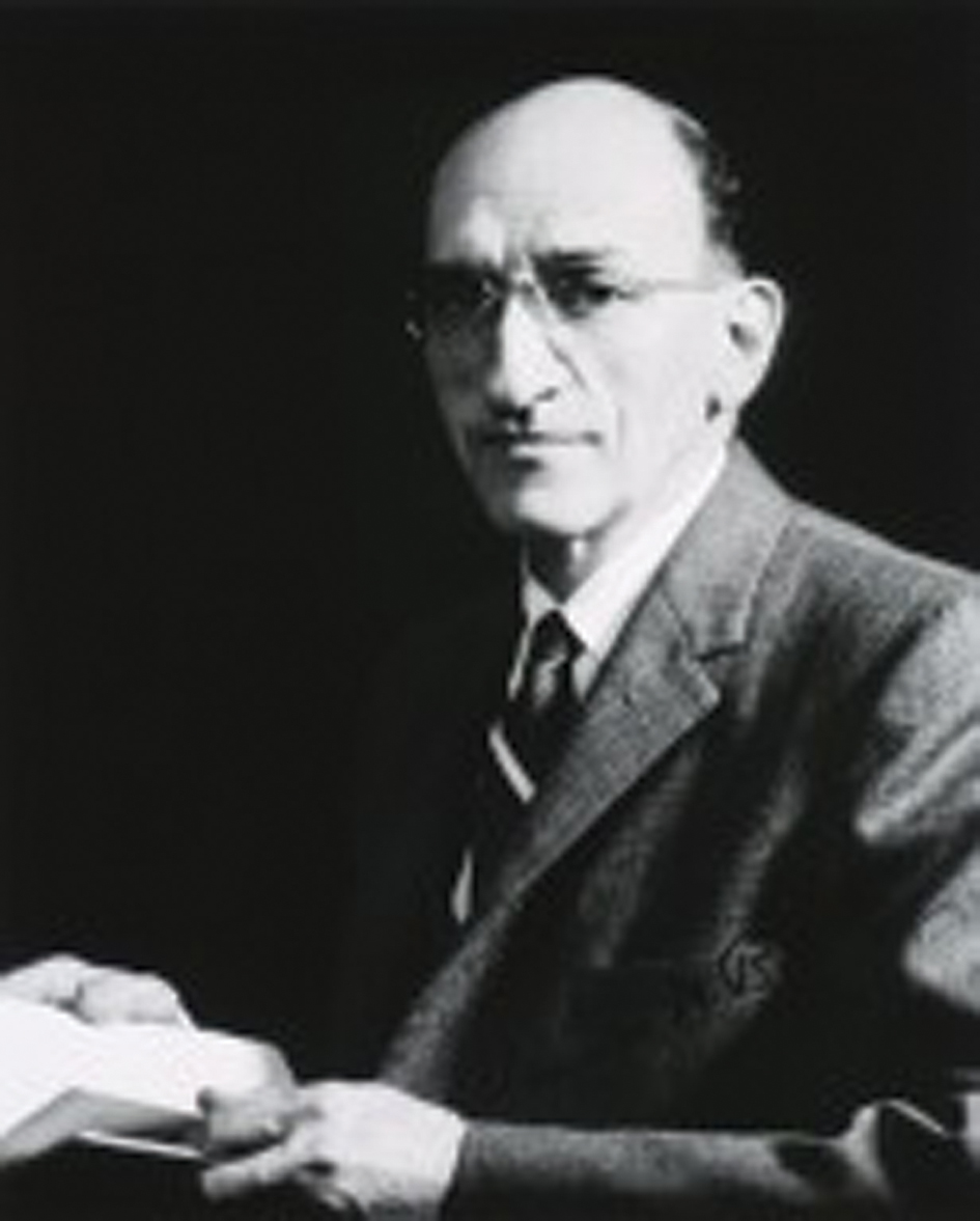 Figure 8. Maurice Nathaniel Richter.
A courtesy of National library of Medicine.
|
|
Sézary syndrome or Sézary
disease [23] |
In a series of papers from 1938 to 1949, Albert Sézary (1880-1956) (Fig. 9), a French dermatologist and syphilologist, described erythroderma with cellules monstrueuses (monster cells) in the skin and blood, which is now known as Sézary syndrome or Sézary disease.
Figure 9. Albert Sézary (1880-1956) |

Table I. Selected Eponyms in the literature of cutaneous lymphomas
REFERENCES
1. Berk DR, Cheng A, Lind AC, Bayliss SJ: Burkitt lymphoma with cutaneous involvement. Dermatol Online J. 2008;28;14:14.
2. Oeppen RS: Denis Parsons Burkitt (1911-1993). Br J Oral Maxillofac Surg. 2003;41:235.
3. Ziemer M, Bauer HI, Fluhr JW, Kaatz M, Elsner P: Primary cutaneous follicle center lymphoma -’crosti lymphoma’: what can we learn? Am J Clin Dermatol. 2008;9:133-6.
4. Annonymous [Agostino Crosti].: G Ital Dermatol Minerva Dermatol. 1966;107:399-412.
5. Gray Y, Schwartz S: Dutcher bodies in chronic synovitis. Arch Pathol Lab Med. 2002;126:199-201.
6. Dutcher TF, Fahey JL: The histopathology of the macroglobulinemia of Waldenström. J Natl Cancer Inst. 1959;22:887–917.
7. Bain BJ: Dutcher bodies. Am J Hematol. 2009;84:589.
8. Cantwell AR Jr. The Russell Body-The Forgotten Clue to the Bacterial Cause of Cancer. JOIMR. 2003;1:1.
9. Mott FW: Proc R Soc London. 1905;76:235-42.
10. Introcaso CE, Kantor J, Porter DL, Junkins-Hopkins JM: Cutaneous Hodgkin’s disease. J Am Acad Dermatol. 2008;58:295-8.
11. Scully C, Langdon J, Evans J: Marathon of eponyms: 8 Hodgkin disease or lymphoma. Oral Dis. 2010;16:217-8.
12. Bonadonna G: Historical review of Hodgkin’s disease. Br J Haematol. 2000;110:504-11.
13. Parry M: Dorothy Reed Mendenhall (1874-1964). Am J Public Health. 2006;96:789.
14. Schmidt G: [Knowledge of the Austrian pathologist Carl Sternberg (1872-1935). Attempt at a historical presentation of clinical information about lymphogranulomatosis]. Pathologe. 1992;13:296-300.
15. Brzezinski P, Pessoa L, Galvão V, Barja Lopez JM, Adaskevich WP, Niamba PA, et al: Dermatology Eponyms – sign –Lexicon (H). Our Dermatol Online. 2013;4:130-43.
16. Carlesimo M, Tammaro A, Cox C, Mari E, Fidanza L, Narcisi A et al: A Case of Ketron-Goodman Disease. Case Rep Dermatol. 2009;12:1:39-43.
17. Steffen C: Ketron-Goodman disease, Woringer-Kolopp disease, and pagetoid reticulosis. Am J Dermatopathol. 2005;27:68-85.
18. Cribier B. History: Frederic Woringer (1903-1964) and Woringer-Kolopp disease. Am J Dermatopathol. 2005;27:534-45.
19. Summers TA Jr, Rush W, Aguilera N, Lupton G: Cutaneous involvement in the lymphoepithelioid variant of peripheral T-cell lymphoma, unspecified (Lennert lymphoma). Report of a case and review of the literature. J Cutan Pathol. 2009;36:25-30.
20. Klapper W, Koch K, Mechler U, Borck C, Fuhry E, Siebert R: Lymphoma‚ type K.’-in memory of Karl Lennert (1921-2012). Leukemia. 2013;27:519-21.
21. Steffen C: The man behind the eponym: Lucien Marie Pautrier–Pautrier’s microabscess. Am J Dermatopathol. 2003;25:155-8.
22. Yu L, Bandhlish A, Fullen DR, Su LD, Ma L: Cutaneous Richter syndrome: report of 3 cases from one institution. J Am Acad Dermatol. 2012;67:87-93.
23. Steffen C: The man behind the eponym dermatology in historical perspective: Albert Sézary and the Sézary syndrome. Am J Dermatopathol. 2006;28:357-67.
Comments are closed.