Dermatitis herpetiformis Duhring – case report
Zoran Vrucinic
Department of Dermatology, University Hospital Clinical Center, Banja Luka, Bosnia and Herzegovina
Corresponding author: Dr. Zoran Vrucinic, MD PhD, E-mail: teledermatologija@gmail.com
Submission: 15.11.2017; Acceptance: 25.03.2018
How to cite this article: Vrucinic Z. Dermatitis herpetiformis Duhring – case report. Our Dermatol Online. Our Dermatol Online. 2018;9(e):e1.
ABSTRACT
Dermatitis herpetiformis (DH) is rare, persistent an autoimmune itchy skin eruption associated with gluten sensitive disorder. DH is characterized by grouped excoriations, erythematous, urticarial plaques, and papules with vesicles. We present a case of 15th years old young man with first skin lesions started five years ago as erythematous rush on elbows and knees. Our patient had, after few months, eruption erythematous plaques on the skin of the extensor side upper and lower extremities, back, neck and both gluteal region. Therapy: Gluten-free diet, methylprednisolone 40mg i.m. 10 days, then 36mg per os in the morning 5 days, and reduce dose methylprednisolone every 5 days for 4 mg less, Ranitidin tabl 150 mg 2×1,Thopical therapy: Alklometazon ung 2x. After one month, our patient had good regression skin lesion and less pruritus and sistemic therapy with methyprednisolone was excluded, with significant clinical improvement, without new lesions and without DH lesions after six monts.Gluten-free diet is control of the cutaneous manifestations of DH in most patients. Pharmacotherapy used to treat DH are Dapsone and sulfapyridine like the primary medication. Other medication include systemic corticosteroid therapy, colchicine, cyclosporine, and azathioprine.
Key words: Dermatitis herpetiformis; Gluten sensitive disorder; Methylprednisolone
INTRODUCTION
Dermatitis herpetiformis (DH) is rare, persistent an autoimmune itchy skin eruption associated with gluten sensitive disorder. DH is characterized by grouped excoriations, erythematous, urticarial plaques, and papules with vesicles. The classic location for dermatitis herpetiformis lesions is on the extensor surfaces of the elbows, knees, buttocks, and back. DH is also known as Duhring disease or gluten rash [1,2].
CASE REPORT
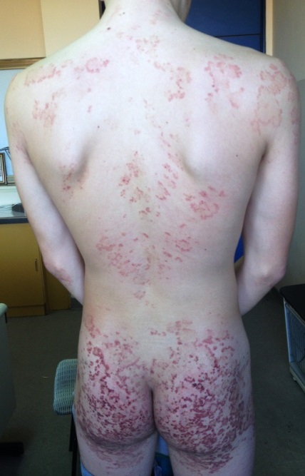
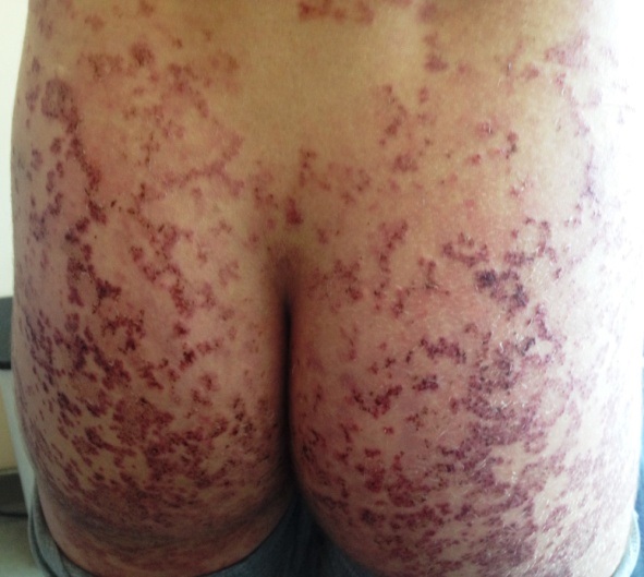
Presentation of 15th years old young man with first skin lesions started five years ago as erythematous rush on elbows and knees. After few months the lesions as confluent erythematous plaques on the skin of the extensor side upper and lower extremities, back, neck and both gluteal region (Figs. 1a and 1b). On the same places thera are few single and linear excoriations and non adherent crust with intensive itching. From first skin lesions till nowadays, patient was treated by several dermatologist under diagnosis of pruritus, atopic dermatitis and eczematous dermatitis and used different types of local and systemic corticosteroids therapy, oral antibiotics, antihistamines and one year of Dapson 50mg a day,without improvement.
On clinical examination, the patient was symmetrically distributed confluent erythematous papules,plaques and crusted lesions on the skin the extensor surface upper and lower extremitas, back, neck and both gluteal region. The nails and theet were normal. He had severe itching.
Laboratory Examinations:
Transglutamine Antibody IgA 113 U/ml (lower limit 10 U/ml), Transglutamine Antibody IgG negative. (05.12.2014).
PHD analysis:
Irregular acanthosis of epidermis. Partially, signs of inter and intra fluid accumulation. Dermal papilla’s elongated and edematous. In single papilla’s are groups of neutrophilic granulocytes (microabscess). Focal, in papilla’s, presens of small splits filled with fibrin and neutrophilic granulocytes. Capillary blood vessels of the upper dermis are dilated, lined with hyperplastic and hypertrophic endothelial cells. Around are lymphocytic and neutrophilic granulocytes.
Skin biopsy:
PHD No 508/03.12.2014. confirmed the diagnosis of Dermatitis Herpetiformis.
Imunology:
IgE 424 kU/l (lower limit 80), Transglutamine Antibody IgA 77,2 U/ml (lower limit 10), Transglutamine Antibody IgG negative, Anti-gliadin Antibodies IgG 14,5 U/ml (lower limit 10), Anti-gliadin Antibodies Ig A -negative (24.11.2015).
Complete blood count:
RBC 4.74 x10¹²/L, Hgb 135 g/l, MCV 86.7, MCH 28.4, Hct 0.41 L/L, WBC 8.09 x10⁹/L, PLT 197×109/L.
Differential diagnosis
The differential diagnosis is concerned with scabies, atopic dermatitis, contact eczema, impetigo and other autoimmunembullous disorders like lineal IgA dermatosis and penfigo. The histopathological findings are fundamental as are the direct immunofluorescence findings [1,2].
Therapy
Gluten-free diet, methylprednisolone 40mg i.m. 10 days, then 36mg per os in the morning 5 days, and reduce dose methylprednisolone every 5 days for 4 mg less, Ranitidin tabl 150 mg 2×1,
Thopical therapy: Alklometazon ung 2x,
Follow-up evaluation
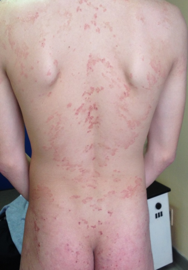
Our patient had good regression skin lesion and less pruritus after one month (Fig. 2).
In January 2016, sistemic therapy with methyprednisolone was excluded.
Topical therapy: 30% fluocinolone acetonide in dermobasae.
Gluten-free diet for life is strongly recommended in our patients with DH.
Patient reports significant clinical improvement without new lesions after two monts (Fig. 3a) and without DH lesions after six monts (Fig. 3b).


International prevalence of DH has been reported as high as 10 cases per 100,000 population. Any age persons may be affected but rare in children [3].
DH is characterized by grouped excoriations, erythematous, urticarial plaques, and papules with vesicles. The classic location for dermatitis herpetiformis lesions is on the extensor surfaces of the elbows, knees, buttocks, and back. Dermatitis herpetiformis lesions are very pruritic. Secondary infection is result from scratching, and requiring antibiotic therapy. Insomnia is very common.
Results of a gluten-free diet is control of the cutaneous manifestations of dermatitis herpetiformis in most patients [4,5]. A gluten-free diet may reduce the incidence of dermatitis herpetiformis–associated lymphomas [6].
Skin biopsy is necessary for diagnosis of DH. DIF is the gold standard for the diagnosis of DH [7-9,11].
Pharmacotherapy used to treat DH are Dapsone (diaminodiphenyl sulfone) and sulfapyridine like the primary medication. Patients should be monitored for the adverse effects of dapsone, primarily hemolytic anemia, methemoglobinemia, agranulocytosis, and neuropathy. Other treatments for dermatitis herpetiformis include systemic corticosteroid therapy, colchicine, cyclosporine, and azathioprine [7].
Patients with DH should be evaluated at regular intervals: 6 months after diagnosis and then yearly [10].
REFERENCES
1. Duhring L. Dermatitis herpetiformis. JAMA. 1884;3:225.
2. Sardy M, Karpati S, Merkl B, Paulsson M, Smyth N. Epidermal transglutaminase (TGase 3) is the autoantigen of dermatitis herpetiformis. J Exp Med. 2002;195:747-57.
3. Templet JT, Welsh JP, Cusack CA. Childhood dermatitis herpetiformis: a case report and review of the literature. Cutis. 2007;80:473-6.
4. Ciacci C, Ciclitira P, Hadjivassiliou M, Kaukinen K, Ludvigsson JF, McGough N, et al. The gluten-free diet and its current application in coeliac disease and dermatitis herpetiformis. United European Gastroenterol J. 2015;3:121-35.
5. Kadunce DP, McMurry MP, Avots-Avotins A, Chandler JP, Meyer LJ, Zone JJ. The effect of an elemental diet with and without gluten on disease activity in dermatitis herpetiformis. J Invest Dermatol. 1991;97:175-82.
6. Sigurgeirsson B, Agnarsson BA, Lindelöf B. Risk of lymphoma in patients with dermatitis herpetiformis. BMJ. 1994;308:13-5.
7. Smith JB, Taylor TB, Zone JJ. The site of blister formation in dermatitis herpetiformis is within the lamina lucida. J Am Acad Dermatol. 1992;27:209-13.
8. Caproni M, Antiga E, Melani L, Fabbri P. The Italian Group for Cutaneous Immunopathology Guidelines for the diagnosis and treatment of dermatitis herpetiformis. J Eur Acad Dermatol Venereol. 2009;23:633–8.
9.Fry L. Dermatitis herpetiformis: problems, progress and prospects. Eur J Dermatol. 2002;12:523–31.
10. Kotze LM. Dermatitis herpetiformis, the celiac disease of the skin! Arq Gastroenterol. 2013;50:231–5.
11. Huber C, Trüeb RM, French LE, Hafner J. Negative direct immunofluorescence and nonspecific histology do not exclude the diagnosis of dermatitis herpetiformis Duhring. Int J Dermatol. 2013;52:248–9.
Notes
Source of Support: Nil,
Conflict of Interest: None declared.
Comments are closed.