A case report of small-plaque parapsoriasis
Katarzyna Borowska 1,2, Bartłomiej Zoń3, Jan Faryna4
1Department of Histology and Embryology with Experimental Cytology Unit, Medical University of Lublin, Lublin, Poland, 2Medical Center CADERM in Warsaw, Poland, 3Clinical Department of Plastic, Reconstructive Surgery and Burns Treatment, Military Medical Institute, Warsaw, Poland, 4Department of Patholomorphology and Cytology Bielanski Hospital Warsaw, Poland
Corresponding author: Prof. Katarzyna Borowska, MD PhD
How to cite this article: Borowska K, Zoń B, Faryna J. A case report of small-plaque parapsoriasis. Our Dermatol Online. 2022;13(e):e48.
Submission: 30.05.2022; Acceptance: 05.06.2022
DOI: 10.7241/ourd.2022e.48
Citation tools:

Copyright information
© Our Dermatology Online 2022. No commercial re-use. See rights and permissions. Published by Our Dermatology Online.
ABSTRACT
Small-Plaque Parapsoriasis (SPP) is a relatively rare, chronic, idiopathic dermatosis, most often seen in middle age people. This disease shows a definite male predominance of approximately 3-4: 1. It is characterized by the presence of round or oval erythematous, slightly scaly plaques on the limbs and trunk. The plaques are usually asymptomatic. The etiology of parapsoriasis is still unknown. This report describes the case of a 64-year-old man with SPP. Histopathologic examination of the skin specimen was compatible with clinical diagnosis.
Key words: Small-Plaque Parapsoriasis; SPP
INTRODUCTION
Parapsoriasis en plaque describes a group of clinically variable inflammatory diseases that can be characterized by erythematous and well-demarcated, slightly scaly plaques and patches on the trunk and/or proximal extremities. The plaques are usually asymptomatic. The etiology of parapsoriasis is still unknown [1]. They are classified into two main groups: Small-Plaque Parapsoriasis (SPP) and Large-Plaque Parapsoriasis (LPP). SPP is defined by lesions < 5 cm in diameter, lesions in LPP > 5 cm in diameter. SPP extremely rarely transforms into Mycosis Fungoides (MF). If SPP is widely known as benign, the main focus is on the distinction of LPP as a “benign inflammatory dermatosis” from an incipient form of MF [2]. In this article we describe the case of a patient suffering from SPP.
CASE REPORT
A 64-year-old male patient presented to Medical Center CADERM in Warsaw with long persisting erythematous patches on his upper and lower limbs. Plaques were of oval and round shape, pretty well marginated. They were of light red colour, covered with fine scales with a slightly wrinkled surface (Figs. 1 – 2). The number of patches has been increasing for one year and then it has remained relatively constant. At time, the patient sometimes complains of itching.
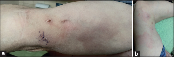 |
Figure 1: (a and b). Erythematous plaques on the right upper limbs area. |
 |
Figure 2: Erythematous plaques on the right lower limbs area. |
A skin segment of approx. 1 x 0.5 cm. was collected under local infiltrative anesthesia (2% xylocaine) from a right arm lesion. The wound after procedure was sutured with simple interrupted DAFILON 5/0 skin sutures. The sutures were removed 7 days after the procedure – the wound healed by primam without any complications. The biopsied specimens were initially processed with routine histological technique, the slides being stained with Hematoxylin and Eosin. Histopathologic examination of the skin specimen revealed epidermal atrophy, focal parakeratosis, perivascular dermal infiltrate of mononuclear cells with exocytosis in the epidermis.
The patient’s medical history also included hypertension, diabetes, thyroiditis. The plaques have been remarkably stubborn, no responding to treatment with steroid creams, therefore, phototherapy treatment is scheduled.
DISCUSSION
SPP is a relatively rare, chronic, idiopathic dermatosis, most often seen in middle age people. This disease shows a definite male predominance of approximately 3-4: 1. It is characterized by the presence of round or oval erythematous, slightly scaly plaques on the limbs and trunk, which histologically reveal mild eczematous changes. Histopathologic examination of the skin specimen revealed epidermal atrophy, focal parakeratosis, perivascular dermal infiltrate of mononuclear cells with exocytosis in the epidermis [3]. This finding was compatible with our clinical diagnosis. The histological sections of LPP describe superficial lymphocyte infiltration with different degrees of epidermotropism, with more than 30% of the cases being described to develop into MF [4]. LLP is difficult to differentiate from early Cutaneous T-cell Lymphoma (CTCL) by clinical features, histopathological characteristics or immunophenotype [5]. There is no marker to identify cases prone to progression. Both subtypes (SPP and LPP) may remain indolent for many years. Pityriasis rotunda should also be taken into account in clinical differentiation from LPP [6].
Occasionally patients with the clinical and pathologic presentation of SPP may develop typical symptoms of MF. Philips CA et al. described branch duct-type intraductal papillary mucinous neoplasm presenting as paraneoplastic SPP [7]. Treatment of SPP is usually unnecessary but most often includes emollients, topical corticosteroids and phototherapy [8,9].
CONCLUSION
This case deserves a very long clinical and histological assessment. Periodic clinical follow-up and biopsies give the best indication of potential risk of developing CTLC, which is very rare.
Consent
The examination of the patient was conducted according to the principles of the Declaration of Helsinki.
The authors certify that they have obtained all appropriate patient consent forms, in which the patients gave their consent for images and other clinical information to be included in the journal. The patients understand that their names and initials will not be published and due effort will be made to conceal their identity, but that anonymity cannot be guaranteed.
REFERENCES
1. Belousova I, Vanecek T, Samtsov AV, Michal M, Kazakov D. A patient with clinicopathologic features of small plaque parapsoriasis presenting later with plaque-stage mycosis fungoides:report of a case and comparative retrospective study of 27 cases of “nonprogressive“small plaque parapsoriasis. J Am Acad Dermatol. 2008;59:474-82.
2. Gug G, Solovan C. From benign inflammatory dermatosis to cutaneous lymphoma. dna copy number imbalances in mycosis fungoides versus large plaque parapsoriasis. Medicina (Kaunas). 2021;57:502.
3. Baderca F, Chiticariu E, Baudis M, Solovan C. Biopsying parapsoriasis:Quo vadis. Are morphological stains enough or are ancillary tests needed?Rom J Morphol Embryol. 2014;55:1085–92.
4. Fujii K, Kanekura T. Next-Generation sequencing technologies for early-stage cutaneous T-Cell Lymphoma. Front Med. 2019;6:181.
5. Kikuchi A, Naka W, Harada T, Sakuraoka K, Harada R, Nishikawa T. Parapsoriasis en plaques:its potential for progression to malignant lymphoma. J Am Acad Dermatol. 1993;29:419–22.
6. El Kadiri S, Bay Bay H, Chaoui R, Douhi Z, Elloudi S, Mernissi FZ. Pityriasis rotunda:A case from Morocco. Our Dermatol Online. 2020;11(e):e51.1-e51.2.
7. Philips CA, Augustine P, Kumar L, Joseph G, Mahadevan P. Branch duct-type intraductal papillary mucinous neoplasm presenting as paraneoplastic small plaque para-psoriasis. Indian Dermatol Online J. 2018;9:40-3.
8. Branisteanu DE, Dirzu DS, Toader MP, Branisteanu DC, Nicolescu AC, Brihan I, et al. Phototherapy in dermatological maladies (Review). Exp Ther Med. 2022;23:259.
9. Duarte IA, Korkes KL, Amorim VA, Kobata C, Buense R, Lazzarini R. An evaluation of the treatment of parapsoriasis with phototherapy. An Bras Dermatol. 2013;88:306-8.
Notes
Source of Support: Nil,
Conflict of Interest: None declared.
Request permissions
If you wish to reuse any or all of this article please use the e-mail (brzezoo77@yahoo.com) to contact with publisher.
| Related Articles | Search Authors in |
|
|
 http://orcid.org/0000-0003-0964-6169 http://orcid.org/0000-0003-0964-6169 |

Comments are closed.