Dermoscopy of palmoplantar keratoderma: development and analysis methods
Ryme Dassouli , Sara Elloudi, Souad Choukri, Hanane Baybay, Zakia Douhi, Fatima Zahra Mernissi
Department of Dermatology, University Hospital Hassan II Fès, Morocco
Corresponding author: Ryme Dassouli, MD
How to cite this article: Dassouli R, Elloudi S, Choukri S, Baybay H, Douhi Z, Mernissi FZ. Dermoscopy of palmoplantar keratoderma: development and analysis methods. Our Dermatol Online. 2022;13(e):e44.
Submission: 19.01.2022; Acceptance: 18.04.2022
DOI: 10.7241/ourd.2022e.44
Citation tools:

Copyright information
© Our Dermatology Online 2022. No commercial re-use. See rights and permissions. Published by Our Dermatology Online.
ABSTRACT
Palmoplantar keratoderma (KPP) is a group of inherited or acquired conditions characterized by palmar and plantar hyperkeratosis. The analysis can be difficult, however, thanks to dermoscopy, which is a non-invasive means of exploration, certain signs are very orienting for the right diagnosis. The available data concerning the dermoscopic profile of palmoplanar pathologies are still limited in the literature. we have reviewed the dermoscopic signs of KPP which target the acquired inflammatory dermatoses frequently found in the routine consultation of the dermatologist. The aim is to realize a dermoscopic guide for the dermatologist in front of KPP and to facilitate the diagnostic orientation in front of this topography using the Dermoscope. We have illustrated with dermoscopic images taken from patients followed in consultation.The analyzed images were taken with minimal pressure to preserve vessel morphology, and immersion with a clear, thick, colorless gel to ensure better visualization.
Key words: Dermoscopy; Eczema; Palmoplantar; Psoriasis; Lichen; Syphilides
INTRODUCTION
Palmoplantar keratoderma is a thickening of the stratum corneum of the palms and soles. Among these hyperkeratosis, 3 groups can be distinguished: hereditary KPP, genodermatoses with KPP and acquired KPP [1].
Acquired KPPs encompass inflammatory, infectious or tumoral pathologies affecting the palms and soles and responsible for hyperkeratosis. The clinical distinction between the different keraoderma is generally difficult, requiring anatomopathological confirmation [2].
Psoriasis (PP), Contact Dermatitis (DC), Lichen Planus (LP), Lichen Nitidus (LV), Pytiriasis Rubra Pilaire (PRP) are common inflammatory diseases of the skin. Mycosis fungoides (MF) is a non-melanocytic tumoral entity that can also be responsible for palmoplantar keratoderma. Their characteristic appearance allows clinical diagnosis in a high proportion of patients. However, palmoplantar presentations sometimes exist and can lead to difficulties in differentiating between these entities. In these cases, histopathology contributes significantly to the accurate diagnosis [3].
Indeed, dermoscopy is a rapid and non-invasive technique that offers the advantage of evaluating pigmented and vascular structures that are not clinically visible. This must-have method for the dermatologist provides additional information at a sub-macroscopic level that can help the dermatologist differentiate between two or more conditions that are barely recognizable to the naked eye [4,5].
This narrative review is based on literature searches in the PubMed online database. Key articles were identified, including recent systematic reviews and guidelines from different societies. For some key articles, a search of references and cited articles was performed to identify other relevant articles.
DERMOSCOPIC EVALUATION METHODOLOGY OF A PALMOPLANTAR AREA
Palmoplantar anatomy
The palmo-plantar skin is marked by deep structure in ordered relief: dermatoglyphs with formation of ridges and furrows. The ridges represent the peaks of the mountains, while the furrows correspond to the valleys between its mountains. The ridges are much wider and not interspersed with the orifices of the sweat ducts.
ANALYSIS OF KERATODERMA
Palmar keratoderma is a heterogeneous and frequent entity encompassing etiologies. There are no reliable morphological characteristics that would allow to distinguish between the different dermatitis of the hand [6]. The analysis of keratoderma is based on a good analysis of the clinical and morphological model, on the dermoscopic description and on the histological confirmation of the diagnosis.
In recent years, dermoscopy has proven to be a useful tool to aid in the non-invasive diagnosis of various general dermatological disorders. The most important criteria to consider when using dermoscopy in general dermatology are: (i) morphology/arrangement of vascular structures; (ii) scaly patterns; (iii) colors; (iv) follicular abnormalities; and (v) specific features (clues) [6–8], inflammatory skin conditions, including palmoplantar topography [5,8].
SEMIOLOGICAL ELEMENTS OF PALMOPLANTAR DERMOSCOPY
-
a-Background color:
-
Can take on a light red color (Fig. 1a) dull red (Fig. 1b) or a yellowish color (Fig. 1c)
-
b-The morphology of vascular structures:
-
We come across vascular structures in points (Fig. 1d) characterized by a size not exceeding 1mm, globular vascular structures (Fig. 1e) or glomerular (Fig. 1f) linear vessels or hairpins.
-
c-The distribution of vessels:
can be homogeneous, symmetrical, uneven asymmetrical, patchy or along the epidermal ridges. The keratinocyte thickness of the palmolantar region can sometimes mask the vascular structures during an inflammatory pathology, one then speaks of an undifferentiated structure (Fig. 1g).
-
d-Scales:
Can take on a whitish (Fig. 1h), yellowish (Fig. 1i) or yellowish-white (Fig. 1j) color.
-
e-The distribution of scales:
It also has an important semiological value, it can be asymmetrical (Fig. 2a) take a peripheral disposition (Fig. 2b), diffuse homogeneous on all the lesion (Fig. 2c), a central concentrated distribution (Fig. 2d) or else distributed along the along the furrows (Fig. 2e).
-
f-The cells:
The presence of orange-brown dots/globules (Fig. 2f) corresponding to tiny spongiotic vesicles (not visible to the naked eye) resistant to rupture due to the increased thickness of the keratin layer at these sites. Globules with a pale center and dark peripheral edges correspond to central vesicles surrounded by hyperkeratosis.
One can also find other structures such as areas without yellowish or orange structures.
 |
Figure 1: (a) Dermoscopic image showing a light red background. (b) Dermoscopic image showing a dull red background. (c) Dermoscopic image showing a yellowish background. d) Vascular structures in points. (e) Red globular vascular structures. (f) Glomerular vascular structures. (g) Undifferentiated vascular structure (masked by the thickness of the keratinocytes). (h) Whitish scales. (i) Yellowish scales. (j) Yellowish white scales. |
 |
Figure 2: (a) Asymmetric distribution of scales on the lesion. (b) Peripheral arrangement of scales on the lesion. (c) Diffuse homogeneous distribution of scales on the whole lesion. d) A central concentrated distribution. (e) Distribution of scales along the furrows. (f) Presence of orange globules (->), orange globules surrounded by a greyish halo (->) |
PALMOPLANTAR PSORIASIS
Psoriasis is a chronic, relapsing inflammatory disease that triggers the appearance of scaly red lesions on the skin. The most common type of psoriasis is psoriasis vulgaris, which accounts for 90% of cases [4] Almoplantar psoriasis is characterized by different morphological patterns, including the well-defined erythematous border (Fig. 3a), fissures, pustular lesions and hyperkeratotic plaques. Its prevalence can reach 4% of all cases [4,5] Palmoplantar psoriasis can be a diagnostic challenge because it is difficult to differentiate it from eczematous entities such as dyshidrotic eczema, contact dermatitis, pompholyx, palmoplantar pustulosis, acquired keratoderma, pityriasis rubra pilaris, and tinea manuum or paedis [4].
 |
Figure 3: (a) Fissured psoriatic plantar keratoderma with well-limited erythematous borders. (b) Dull red background with asymmetrically distributed whitish scales in psoriatic KPP. (c) Dot-like and glomerular vessels with homogeneous distribution on a light red background in psoriatic KPP. (d) Dermoscopy revealing white esquamia surrounding desquamative areas and a flower-like pattern on healthy skin in psoriatic KPP [4]. (e) Dermoscopic image of cPP eczema showing orange-brown globules and globules with a pale center and a dark peripheral border, orange-yellow scales on a dull red background. (f) Dermoscopic image of PP eczema showing patchy vessels and fine white-yellowish scales on a light red background. |
Dermoscopy of plaque psoriasis usually shows a characteristic pattern consisting of diffuse, symmetrical, regular white scales, dotted vessels distributed over a light or dull red background (Fig. 3b) [5–8]. Lallas et al. reported that the combination of regularly distributed dotted vessels (Fig. 3c) on a light red background associated with diffuse white scales were highly predictive of plaque psoriasis and allowed a correct diagnosis with 88% specificity and 84.9% sensitivity [6,9]. Annular globular vessels are rare but have a high predictive value for psoriasis [5,10]. The scales can take different distributions, they can be homogeneous, with a peripheral arrangement, along the furrows or with a concentrated central distribution.
Martinez and AL [4] had described a new dermoscopic description of PP showing a flower-like configuration in areas without desquamation. Histopathologically, these areas may correspond to the formation of spongiotic vesicles and the white linear structures represent areas of partially preserved keratinocytes (Fig. 3d).
PALMO-PLANTAR ECZEMA
Hyperkeratotic eczema is characterized by painful hyperkeratotic patches, including scales, sores and cracks. Usually the palms and volar surfaces of the fingers are involved, and sometimes also the plantar surfaces of the feet. Although the exact etiology is not known, atopy, contact allergy, irritation and friction could be responsible [5,6]. Differential diagnosis between hyperkeratotic palmoplantar eczema and isolated PPP can be difficult. [6–8].
The most specific dermoscopic features of chronic hand eczema include orange-brown dots/globules that correspond to palmoplantar spongiotic vesicles, yellowish scales, and yellow-orange crusts (Fig. 3e). Punctate vessels are indicative of eczema when seen in an uneven distribution with yellow scales (Figs. 3f and 1j) [5,9]. Globules with a pale center and a dark peripheral rim are also described. The dark peripheral rim may be associated with hyperkeratotic foci around the vesicles, but this is not significant for the differential diagnosis with psoriasis [5,10].
Punctate vessels are indicative of eczema when seen in an uneven distribution with yellow scales [5,10–13] The yellow scales correspond to hyperkeratosis mixed with dried serous fluids.
The table below summarizes a comparison between dermoscopic models of PP eczema VS palmoplantar psoriasis (Table 1).
 |
Table 1: Comparative table of dermoscopic signs of psoriasis and eczema. |
PYTIRIASIS RUBRA PILARIS
Pityriasis rubra pilaris (PRP) is a relatively rare skin disease, characterized clinically by keratotic follicular plugs, red to orange plaques and palmoplantar hyperkeratosis (Fig. 4a) [9,14]. However, in cases with an atypical clinical presentation, PRP is sometimes confused with psoriasis, both clinically and histopathologically [14].
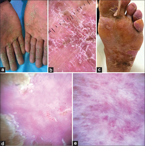 |
Figure 4: (a) Acquired palmar keratoderma due to pityriasis rubra pilaris [13]. (b) Dermoscopic examination shows whitish scales and homogeneous orange areas, without structure, distributed in places and of different sizes (black arrows) [13]. (c) Palmar fissure keratoderma acquired due to mycosis fungoides [13]. (d) Dermoscopy shows relatively large amber scales (black arrows) on a white to pinkish background; scattered whitish scales and several non-specific reddish fissures are also present [13]. (e) Dermoscopic image showing linear short fine vessels «short wavy vessels» on a pinkish white background in a palmar keratoderma of mycosis fungoides. |
Dermoscopic shows whitish scales and homogeneous orange areas, without structure, unevenly distributed and of different size [14,15], uneven fine whitish scales (Fig. 4b). The vascular pattern is undifferentiated because masked by the keratinocyte thickness [15].
MYCOSIS FUNGOIDES PP
Mycosis fungoides (MF) is one of the primary cutaneous lymphomas that initially manifests in the skin and may include secondary extracutaneous invasion [16]. Lesions can take different forms: Annular plaques, hyper-keratotic plaques (Fig. 4c), verrucous nodules, dyshidrotic pseudo-eczema or even pustulosis. Palmoplantar location remains very rare and diagnosis is difficult when palmoplantar involvement is isolated.
The dermoscopic signs found are: a white to pinkish background color, relatively large amber scales, relatively large amber scales (black arrows) on a white to pinkish background; scattered whitish scales and several non-specific reddish cracks are also present (Fig. 4d) [16,17]. The large amber scales and a pale background in AK related to mycosis fungoides could be due to the marked/compact hyperkeratosis/acanthosis, which can be observed in this disease. Although the vascular pattern is often masked by the thickness of the stratum corneum, several vascular structures can be found, such as fine linear short vessels “corrugated short vessels” [17,18] (Fig. 4e).
PALMAR LICHEN PLANUS
Palmoplantar lichen planus (LPPP) is a localized and infrequent variant of lichen planus (LP). The erythematous and scaly form is the most common, and other clinical presentations include vesicle and petechiae-like lesions, which are rare variants [9,19]. In plantar cases, these lesions are often located on the internal arch, often without involvement of the palms or fingers (Fig. 5a) [19,20]. Lesions on the hands are observed on the lateral edges of the fingers and on the surface of the hand; however, they are less likely to affect the fingertips. Generally an extension towards the rest of the body occurs after plantar expectation [20,21]. The lesions usually resolve on their own within a few months [21].
 |
Figure 5: (a): Plantar keratoderma of a lichen planus made of yellowish keratotic lesions located on the inner arch of the sole of the feet. (b) Dermoscopic image showing concentric yellowish oval areas on a purplish background in plantar keratoderma of lichen planus. (c) Palmar lichen nitidus with hyperkeratotic papules and erythematous plaques. (d) Well-defined oval depressions surrounded by ring-shaped silvery white scales. (e) Mealy plantar keratoderma in dermatophytosis. (f) Whitish scales following the path of focally accentuated furrows (sites of maximum dermatophytic activity), resting on an erythematous background in dermatophytic plantar keratoderma. |
Dermoscopic examination of a case of palmar lichen planus shows rounded yellowish areas, some of which have peripheral star-shaped projections on a purplish background (Fig. 5b) [19,21].
LICHEN NITIDUS
LN is an uncommon lichenoid dermatosis that can be defined as multiple, separate, shiny, pinpoint-shaped, pale skin-colored papules, 1-2 mm in diameter. LN usually affects children and young adults without any predilection for sex. The etiology of the disease is uncertain [21]. The most commonly affected sites are the flexor areas of the upper extremities, the backs of the hands, the trunk and the genitals [22]. Cases of LN localized only on the palms and presenting as hyperkeratotic papules and plaques have been rarely reported (Fig. 5c) [23]. It is difficult to make a diagnosis of palmar LN in the absence of lesions elsewhere on the body. There are certain defined dermoscopic characteristics for LNs.
In the palmoplantar variant, parallel thick linear scales discontinuous by well-defined oval depressions and surrounded by ring-shaped silvery-white scales have been described [23,24]. The axis of the depressed pattern and that of the linear scales are parallel [23,25] (Fig. 5d).
TINEA MANUM
Tinea manuum (TM) is a dermatophyte infection affecting the palm of the hand and the interdigital spaces, which typically presents as mealy scales with hyperkeratoses (keratoderma) and diffuse white scales, with or without mild itching [1,26] (Fig. 5g). Although unilateral or asymmetric involvement and concurrent infection of other sites (particularly the nails and feet) may be useful in aiding the diagnosis of TM, its distinction from similar inflammatory dermatoses is often difficult, resulting in diagnostic delays, errors and unnecessary therapies [26,27].
We find whitish scales mainly located in the furrows [1,27]. they could result from the predilection of dermatophytes to proliferate in a humid environment, such as the palmar furrows [27]. The fundus is often erythematous (Fig. 5h). In the dishydrosic forms one can find brownish scales corresponding to the vesicles embedded between the keratinocytes [28].
PALMOPLANTAR SYPHILIDES
Secondary syphilis occurs following blood or lymphatic dissemination of Treponema. It is characterized by 2 phases: the 1st flowering and the 2nd flowering. At the 2nd flowering, asymptomatic lesions called syphilides settle on the body, trunk, face and limbs then disappear in 10 days without leaving scars. The palmoplantar localization of syphilides is rare but very characteristic of the disease (Fig. 6a and 6b). Sometimes the clinical aspect takes other forms that can make the diagnosis difficult. Progressive dermoscopy finds its place for early identification of palmar syphilides compared to other palmar inflammatory pathologies [29,30].
 |
Figure 6: (a) Syphilis of the palmar surfaces of both hands made of coppery papules surrounded by a Biett’s collar. (b) Rounded coppery red papules surrounded by a Biett’s collar on the palmar surface of the left hand in a syphilitic subject. (c) Dermoscopic image of palmar syphilis showing a thin circular rim progressing outwards (called Biett’s ruff) and surrounding an erythematous lesion with a yellow-orange center. (d) Dermoscopic image showing progressive outward extension of Biett’s ruff and a remarkable attenuation of the erythema and orange background is observed on a healing syphilis. |
The main dermoscopic signs specific to palmar syphilides are Biett’s collarette in the form of a thin circular edge progressing outwards and surrounded by an erythematous halo, the orange-yellow background which corresponds to extravasation of hemosiderin, the pattern vascular remains undefined. Progressive extension of Biett’s collarette outward until disappearance with remarkable attenuation of erythema and orange background is observed in more advanced stages [30] (Fig. 6c and 6d).
CONCLUSION
This article provides a discussion of the different dermoscopic patterns and characteristics of palmoplantar keratoderma. The objective is to develop the dermoscopic characteristics associated with various inflammatory pathologies such as psorias, eczema PP, lichen, syphilis, lichen nitidus, pytiriasis rubra pilaris and mycosis fungoide. allowing to better reason in front of a keratoderma using the dermoscpe. Dermoscopy has been proven to be effective in the diagnostic guidance of KPP. However, more studies should be done to define the specific dermoscopic characteristics of the palmoplantar topography of the different pathologies and to better differentiate between diagnoses using the Dermoscope.
REFERENCES
1. Thomas BR, O’Toole EA. Diagnosis and Management of Inherited Palmoplantar Keratodermas. Acta Derm Venereol. 2020;100:0094.
2. Errichetti E, Stinco G. Dermoscopy in General Dermatology:A Practical Overview. Dermatol Ther (Heidelb). 2016;6:471-507.
3. Çetinarslan T, Türel Ermertcan A, Temiz P. Dermoscopic clues of palmoplantar hyperkeratotic eczema and palmoplantar psoriasis:A prospective, comparative study of 90 patients. J Dermatol. 2020;47:1157-65.
4. Martinez-Rico JC, Gomez-Flores M, Ocampo-Candiani J, Villarreal-Martinez A, Chavez-Alvarez S. New dermoscopic finding for palmoplantar psoriasis:flowers. Australas J Dermatol. 2020;61:257-8.
5. Antonov D, Schliemann S, Elsner P. Hand dermatitis:a review of clinical features, prevention and treatment. Am J Clin Dermatol. 2015;16:257-70.
6. Lallas A, Kyrgidis A, Tzellos TG, Apalla Z, Karakyriou E, Karatolias A, et al. Accuracy of dermoscopic criteria for the diagnosis of psoriasis, dermatitis, lichen planus and pityriasis rosea. Br J Dermatol. 2012;166:1198-205.
7. Lallas A, Apalla Z, Argenziano G, Sotiriou E, Di Lernia V, Moscarella E, et al. Dermoscopic pattern of psoriatic lesions on specific body sites. Dermatology. 2014;228:250-4.
8. Cook LC, Hanna C, Foulke GT, Seiverling EV. Dermoscopy in the Diagnosis of Inflammatory Dermatoses:Systematic Review Findings Reported for Psoriasis, Lupus, and Lichen Planus. J Clin Aesthet Dermatol. 2018;11:41-2.
9. Adabala SS, Doshi BR, Manjunathswamy BS. A Cross-sectional study to assess the role of dermoscopy in differentiating palmar psoriasis, chronic hand eczema, and eczema in psoriatico. Indian Dermatol Online J. 2022;13:78-5.
10. Errichetti E, Stinco G. Dermoscopy in differential diagnosis of palmar psoriasis and chronic hand eczema. J Dermatol. 2016;43:423-5.
11. Yu X, Wei G, Shao C, Zhu M, Sun S, Zhang X. Analysis of dermoscopic characteristic for the differential diagnosis of palmoplantar psoriasis and palmoplantar eczema. Medicine (Baltimore). 2021;100:e23828.
12. Vilas Boas da Silva PT, Rodríguez-Lomba E, Avilés-Izquierdo JA, Ciudad-Blanco C, Suárez-Fernández R. Dermoscopic features of circumscribed palmar hypokeratosis. JAMA Dermatol. 2017;153:609-11.
13. Jha AK, Lallas A, Sonthalia S, Jhakar D, Udayan UK, Chaudhary RKP. Differentiation of pityriasis rubra pilaris from plaque psoriasis by dermoscopy. Dermatol Pract Concept. 2018;8:299-302.
14. Errichetti E, Stinco G. Dermoscopy as a supportive instrument in the differentiation of the main types of acquired keratoderma due to dermatological disorders. J Eur Acad Dermatol Venereol. 2016;30:e229-e231.
15. Magro CM, Nguyen GH. Keratoderma-like T cell dyscrasia:A report of 13 cases and its distinction from mycosis fungoides palmaris et plantaris. Indian J Dermatol Venereol Leprol. 2016;82:395-403.
16. Bilgic SA, Cicek D, Demir B. Dermoscopy in differential diagnosis of inflammatory dermatoses and mycosis fungoides. Int J Dermatol. 2020;59:843-50.
17. – Bombonato C, Pampena R, Lallas A, Giovanni P, Longo C. Dermoscopy of Lymphomas and Pseudolymphomas. Dermatol Clin. 2018;36:377-88.
18. Abreu Velez AM, Howard MS, Pereyo N. Palmar and plantar lichen planus:a case report and review of the literature. An Bras Dermatol. 2015;90:175-7.
19. García-García B, Munguía-Calzada P, Aubán-Pariente J, Argenziano G, Vázquez-López F. Dermoscopy of lichen planus:Vascular and Wickham striae variations in the skin of colour. Australas J Dermatol. 2019;60:301-4.
20. Rieder E, Hale CS, Meehan SA, Leger M. Palmoplantar lichen planus. Dermatol Online J. 2014;20:13030.
21. Jakhar D, Grover C, Kaur I, Sharma S. Dermatoscopic features of lichen nitidus. Pediatr Dermatol. 2018;35:866-7.
22. Taneja N, Mehta N, Arava S, Gupta V. An unusual variant of lichen nitidus:Generalized follicular spinous with perifollicular granulomas. J Cutan Pathol. 2020;47:834-9.
23. Podder I, Mohanty S, Chandra S, Gharami RC. Isolated Palmar Lichen Nitidus-A Diagnostic Challenge:First Case from Eastern India. Indian J Dermatol. 2015;60:308-9.
24. Durusu ?N, Güler D, Gürel G, Yalç?n GŞ. A very rare localization of a rare disease:palmar lichen nitidus. An Bras Dermatol. 2022;97:96-8.
25. Qian G, Wang H, Wu J, Meng Z, Xiao C. Different dermoscopic patterns of palmoplantar and nonpalmoplantar lichen nitidus. J Am Acad Dermatol. 2015;73:101-3.
26. Jakhar D, Kaur I, Sonthalia S. Dermoscopy of tinea manuum. Indian Dermatol Online J. 2019;10:210-1.
27. Errichetti E, Stinco G. Dermoscopy in tinea manuum. An Bras Dermatol. 2018;93:447-8.
28. Errichetti E, Stinco G. Dermoscopy in differential diagnosis of palmar psoriasis and chronic hand eczema. J Dermatol. 2016;43:423-5.
29. Errichetti E, Stinco G. Dermoscopy in differentiating palmar syphiloderm from palmar papular psoriasis. Int J STD AIDS. 2017;28:1461-3.
30. Mathur M, Acharya P, Karki A, Shah J, Kc N. Dermoscopic clues in the skin lesions of secondary syphilis. Clin Case Rep. 2019;7:431-4.
Notes
Source of Support: Nil,
Conflict of Interest: None declared.
Request permissions
If you wish to reuse any or all of this article please use the e-mail (brzezoo77@yahoo.com) to contact with publisher.
| Related Articles | Search Authors in |
|
|
 http://orcid.org/0000-0003-4330-4429 http://orcid.org/0000-0002-5942-441X http://orcid.org/0000-0003-3455-3810 http://orcid.org/0000-0003-4330-4429 http://orcid.org/0000-0002-5942-441X http://orcid.org/0000-0003-3455-3810 |

Comments are closed.