Isolated lip lichen planus mimicking Fordyce hyperplasia
Selma El Kadiri , Hanane Bay Bay, Zakia Douhi, Sara Elloudi, Fatima Zahra Mernissi
Department of Dermatology and Venerology, University Hospital Hassan II, Fez, Morocco
Corresponding author: Selma El Kadiri, MD
How to cite this article: El Kadiri S, Bay Bay H, Douhi Z, Elloudi S, Mernissi FZ. Isolated lip lichen planus mimicking Fordyce hyperplasia. Our Dermatol Online. 2021;12(e):e58.
Submission: 07.03.2021; Acceptance: 27.03.2021
DOI: 10.7241/ourd.2021e.58
Citation tools:

Copyright information
© Our Dermatology Online 2021. No commercial re-use. See rights and permissions. Published by Our Dermatology Online.
Oral lichen planus is a dermatosis that usually affects the buccal mucosa but all the oral structures can be affected [1]. Herein we report a case of a 37-year-old male patient with a 6-month history of multiple raised papules over the upper lip (Fig. 1). Examination of skin,) nails, and buccal mucosa was normal. Polarized light dermoscopy revealed a radial and linear distribution of Wickham striae with scaling and erythematous background (Figs. 2a and 2b). A punch biopsy was performed and revealed hyperkeratosis, vacuolization of the membrane basement, and band-like lymphocytic infiltrate compatible with the diagnosis of lip lichen planus. The patient was treated with dipropionate betamethasone with good improvement.
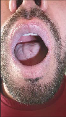 |
Figure 1: Multiple raised papules over the upper lip. |
 ] ] |
Figure 2: (a) Radial distribution of wickham striae with black grey globules, (b) Linear distribution of wickham striae. |
Isolated lip lichen planus is a rare disease that can mimic many conditions such as discoid lip erythematous lupus, allergic cheilitis, Fordyce hyperplasia and Vulgaris pemphigus [1]. Mucoscopy of lip LP has been reported in few observations and recently in a case series of 12 patients. It revealed various patterns of Wickham striae distributed in radial, linear, or leaf venation like pattern. We found a mixed pattern combining radial and linear patterns. It can enhance pigmentary structures such as black grey or brown globules and granules. Vascular structures were also described such as hairpin, linear and dotted vessels [2]. Isolated lip lichen planus must be distinguished from other etiologies of cheilitis and dermoscopy can be the key to achieve this goal.
Consent
The examination of the patient was conducted according to the principles of the Declaration of Helsinki.
The authors certify that they have obtained all appropriate patient consent forms, in which the patients gave their consent for images and other clinical information to be included in the journal. The patients understand that their names and initials will not be published and due effort will be made to conceal their identity, but that anonymity cannot be guaranteed.
REFERENCES
1. Mathur M, Acharya P, Karki A, Kc N, Shah J, Jha A. Isolated lichen planus of lip:Diagnosis and treatment monitoring using dermoscopy. Clin Case Rep. 2018;7:146-8.
2. Neema S, Sandhu S, Kashif AW, Sinha P, Kothari R, Radhakrishnan S. Dermoscopy of lip lichen planus-a descriptive study. Dermatol Pract Concept. 2020;10:e2020076.
Notes
Source of Support: Nil,
Conflict of Interest: None declared.
Request permissions
If you wish to reuse any or all of this article please use the e-mail (brzezoo77@yahoo.com) to contact with publisher.
| Related Articles | Search Authors in |
|
|
 http://orcid.org/0000-0003-3455-3810 http://orcid.org/0000-0002-5942-441X http://orcid.org/0000-0003-3455-3810 http://orcid.org/0000-0002-5942-441X |

Comments are closed.