The value of dermoscopy in the diagnosis of epidermal cyst
Samia Mrabat 1, Zakia Douhi1, Chaymae Jroundi1, Sara Elloudi1, Hanane Baybay1, Fatima Zahra Mernissi1, Amal Douida2, Layla Tahri Elousrouti2, Hinde El Fatemi2
1Department of Dermatology, University Hospital Hassan II, Fes, Morocco, 2Department of Anatomopathology, University Hospital Hassan II, Fe, Morocco
Corresponding author: Samia Mrabat, MD
Submission: 10.09.2020; Acceptance: 10.01.2021
DOI:10.7241/ourd.2021e.17
Cite this article: Mrabat S, Douhi Z, Jroundi C, Eloudi S, Baybay H, Mernissi FZ, Douida A, Elousrouti LT, El Fatemi H. The value of dermoscopy in the diagnosis of epidermal cyst. Our Dermatol Online. 2021;12(e):e17.
Citation tools:

Copyright information
© Our Dermatology Online 2021. No commercial re-use. See rights and permissions. Published by Our Dermatology Online.
ABSTRACT
Epidermal cyst also called epithelial cyst is a very common benign skin tumour. They can appear anywhere on the body but are most common on the face, neck and trunk. They are slow growing and often painless. It is usually easy to diagnose. Sometimes, it can be difficult to make a correct diagnosis for non-typical cases such as multiple, small lesions or those lacking a central comedo-like punctum. We here report the case of an epidermal cyst with no clinically visible punctum in which the dermoscopy was very helpful.
Key words: Dermoscopy; epidermal cyst; skin tumor
INTRODUCTION
An epidermal cyst is a common keratin-filled epithelial-lined cyst. It can generally be diagnosed clinically but a biopsy is required for differential diagnosis with other subcutaneous nodules and for determining the best treatment modality. They present as a roughly circular subcutaneous nodule of variable size, of firm consistency, covered by smooth, normal-colored skin with a central punctum [1]. Although epidermal cysts are the most common cutaneous cysts and frequently encountered in daily dermatologic practice, differential diagnosis of them is very broad and is often a diagnostic challenge. The punctum is a characteristic of epidermal cyst which is a clinically visible orifice representing the orifice of an obstructed hair follicle from which the cyst originates. In case this punctum is not visible, diagnosing epidermal cysts can be difficult [2].
CASE REPORT
A 37-years-old female, with no previous medical history including no local trauma, presented with an asymptomatic nodule on her knee that has been slowly growing over the past three years . Clinical examination found a well limited 2 cm nodule on the right knee of skin color, with firm consistency and smooth surface (Fig.1). The dimple sign was negative and the rest of the dermatological evaluation was normal. Our clinical differential diagnosis included dermatofibroma, pilomatricoma or fibroma. Dermoscopy analysis showed a white-yellow background, telangiectatics vessels, peripheric linear branched vessels and a roughly circular orifice filled with keratin corresponding to the punctum [Fig 2]. This last finding made us think of the possibility of an epidermal cyst which was confirmed by the histology report after the excision of the nodule.
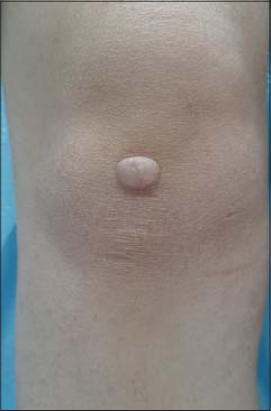 |
Figure 1: Clinical image showing the epidermal cyst of the knee without a clinically visible pore. |
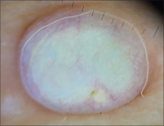 |
Figure 2: Dermoscopic image revealing the presence of the punctum as well as peripheral linear vessels. |
DISCUSSION
Dermoscopy is a non-invasive test without time and spatial limitations, and since recently, polarized dermoscopy has also been used with a cross-polarized lens, which, compared to the conventional non-polarized dermoscopy, allows deeper structures such as blood vessels and collagen to be seen [1]. The diagnostic application of dermoscopy is increasing in general dermatology and for skin tumors because it enables clinicians to visualize morphologic structures that are often invisible to the naked eye. In case of epidermal cysts, the white structure is known to be a dermoscopic clue implying a tumor originating from the hair follicle, and is thought to be a finding histologically appearing corresponding to the keratin mass [4]. Dermoscopic examination may also help visualize the punctum which is a plugged pilosebaceous unit and therefore increase the diagnostic accuracy in a proportion of epidermal cysts with very small puncta [3]. In the absence of a visible pore, the diagnosis of epidermal cyst cannot be excluded, but other differential diagnosis should also be considered [1]. The typical treatment is to remove the cyst after incision, but if the cyst is ruptured and inflamed, or if secondary infection exists, intralesional steroid injection or antibiotic therapy after incision and drainage is required [1]. If the diagnosis of epidermal cyst can be made before surgery, a better treatment option such as surgery with minimal incision and cyst extraction can be chosen; this will produce better cosmetic results [2].
CONCLUSION
Most of the time, the presence of a pore in a cutaneous nodule allows the diagnosis of epidermal cyst to be made. In this case, there was no clinically visible pore but the dermoscopy was of a great help in correcting the diagnosis of epidermal cyst which was later confirmed by the pathology report.
Consent
The examination of the patient was conducted according to the principles of the Declaration of Helsinki.
The authors certify that they have obtained all appropriate patient consent forms, in which the patients gave their consent for images and other clinical information to be included in the journal. The patients understand that their names and initials will not be published and due effort will be made to conceal their identity, but that anonymity cannot be guaranteed.
REFERENCES
1. Suh KS, Kang DY, Park JB, Yang MH, Kim JH, Lee KH, et al. Usefulness of dermoscopy in the differential diagnosis of ruptured and unruptured epidermal cysts. Ann Dermatol. 2017;29:33-8.
2. Mun JH, Park SM, Kim TW, Kim BS, Ko HC, Kim MB. Importance of keen observation for the diagnosis of epidermal cysts:Dermoscopy can be a useful adjuvant tool. J Am Acad Dermatol. 2014;71:e138-40.
3. Zalaudek I, Lallas A, Moscarella E, Longo C, Soyer HP, Argenziano G. The dermatologist’s stethoscope-traditional and new applications of dermoscopy. Dermatol Pract Concept. 2013;3:67-71.
4. Lallas A, Moscarella E, Argenziano G, Longo C, Apalla Z, Ferrara G, et al. Dermoscopy of uncommon skin tumours. Australas J Dermatol. 2014;55:53-62.
Notes
Source of Support: Nil,
Conflict of Interest: None declared.
Request permissions
If you wish to reuse any or all of this article please use the e-mail (brzezoo77@yahoo.com) to contact with publisher.
| Related Articles | Search Authors in |
|
|
 http://orcid.org/0000-0003-3455-3810 http://orcid.org/0000-0003-3455-3810 |

Comments are closed.