A rare case of traumatic panniculitis
Vennela Reddy Sreenivasa , Bittanakurike Narasappav Ragahavendra, Anjan Kumar Patra
Department of Dermatology, MVJ Medical College and Research Hospital, Bangalore, India
Corresponding author: Vennela Reddy Sreenivasa, MD
How to cite this article: Sreenivasa VR, Ragahavendra BN, Patra AK. A rare case of traumatic panniculitis. Our Dermatol Online. Our Dermatol Online. 2021;12(e):e13.
Submission: 04.08.2020; Acceptance: 10.01.2021
DOI: 10.7241/ourd.2021e.13
Citation tools:

Copyright information
© Our Dermatology Online 2021. No commercial re-use. See rights and permissions. Published by Our Dermatology Online.
ABSTRACT
Panniculitis is a rare disorder which usually affects the organ and tissue which are abundant in fat cells. It causes fatty degeneration of the fat cells. Cutaneous panniculitis can be mistaken for other skin and subcutaneous lesions and the diagnosis is usually made histologically. We report a 18 yr old female presented with multiple subcutaneous nodules on right cheek since six months and with history of trauma two years back associated with facial asymmetry and histological examination revealed lobular panniculitis of traumatic etiology.
Key words: Panniculitis; Trauma; Fat Cells
INTRODUCTION
Panniculitis is a rare and poorly understood disorder of adipose tissue. It usually occurs in third to fourth decade of life. It has equal predilection for males and females. Panniculitis is a group of diseases characterized by inflammation of subcutaneous adipose tissue that is the fatty layer under the skin. It is localized to subcutaneous fat and hence termed as panniculitis [1]. In traumatic panniculitis, trauma may be physical including blunt trauma, cold, excessive heat or electrical injury, or chemical injury produced by injection of noxious substances [2]. The clinical picture of traumatic panniculitis is non specific. Panniculitis manifests as more or less well-defined erythematous violaceous nodules or lumps, which coalesce to form indurated erythematous plaques that may be painful [3]. Traumatic panniculitis is usually self limiting disorder and considerable improvement occurs with the time [4].
CASE REPORT
A 18 yr old female presented with darkening of skin on right side of face since 2 years following trauma and multiple raised lesions present over right side of face since 6 months. There were no symptoms as such but for cosmesis. Cutaneous examination revealed a well defined hyperpigmented patch with borders showing four nodules, largest measuring approximately 5×4 cm, mobile, non tender, presented over right cheek with facial asymmetry (Figs. 1 and 2). Clinically differential diagnosis of localized morphea and panniculitis were thought of and skin biopsy was done. Histopathological examination showed epidermis lined by stratified squamous epithelium. Dermis showed extensive mucinous change. Deep dermis showed moderate perivascular chronic inflammatory cell infiltrate composed of lymphocytes. Subcutis showed lobular pattern of panniculitis comprising of lymphohistiocytic infiltrate. All these features were suggestive of lobular panniculitis of traumatic etiology (Fig. 3). X-ray of face was normal.
 |
Figure 1: Hyperpigmented patch with border exhibiting subcutaneous nodules. |
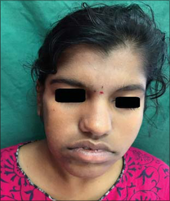 |
Figure 2: Facial asymmetry. |
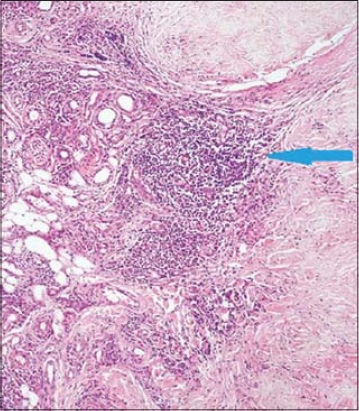 |
Figure 3: Lobular pattern of panniculitis with Lymphohistiocytic infiltrate (blue arrow). |
DISCUSSION
Inflammatory diseases involving the subcutaneous fat comprise a heterogeneous group of disorders named generically panniculitis. These diseases have been classically considered diagnostically challenging both for clinicians and dermatopathologists [4]. Panniculitis classification is performed in accordance with histological examination, the location of the inflammatory process, involvement or not of the blood vessels and the predominant cell type in the infiltrate. If the inflammatory process affects mainly the fibrous trabeculae or septa the panniculitis is called septal, and lobular or lobar when lobes are predominantly affected [3]. Post traumatic panniculitis may present as a solitary or as multiple subcutaneous nodules caused by traumatic separation and consequent devascularization of pieces of subcutaneous fat from its blood supply [4]. The histologic findings are non-specific. They include fat microcysts surrounded by histiocytes, collections of foam cells, and mixed-type of inflammatory cells. Later lesions may develop fibrosis, lipomembranous changes, pseudomembraneous changes, dystrophic calcium deposits or ectopic calcification [5]. Traumatic panniculitis is usually a self-limiting disorder and considerable improvement occurs with time [4].
CONCLUSION
Traumatic panniculitis is rare disorder with a female predominance and needs to be confirmed by skin biopsy. It is usually a self-limiting disorder which requires only symptomatic treatment in most cases.
Consent
The examination of the patient was conducted according to the principles of the Declaration of Helsinki.
The authors certify that they have obtained all appropriate patient consent forms, in which the patients gave their consent for images and other clinical information to be included in the journal. The patients understand that their names and initials will not be published and due effort will be made to conceal their identity, but that anonymity cannot be guaranteed.
REFERENCES
1. Shetty SB, Thomas A, Shetty S, Nair P. A rare presentation of lobular panniculitis in the oral cavity of a 2-year-old patient. J Indian Soc Pedod Prev Dent. 2018;36:324-6.
2. Gupta P, Saikia UN, Arora S, De D, Radotra BD. Panniculitis:A dermatopathologist’s perspective and approach to diagnosis. Indian J Dermatopathol Diagn Dermatol. 2016;3:29-41.
3. Gabriela Martinez Braga, Beatriz Di Martino Ortiz. Septal panniculitis:Clinico-pathological review of the literature and case presentation. Our Dermatol Online. 2014;5:74-82.
4. Requena L. Panniculitis. In:Griffiths CEM, Barker J, Bleiker T, Chalmers R, Creamer D, editors. Rook’s Textbook of Dermatology.9th ed. UK:John Wiley &Sons;2016:99.6-99.52.
5. Wollina U, Heinig B, Langner D, Schmidt N, Hansel G. Posttraumatic panniculitis-Case series and literature review. Glonal Dermatol. 2017;4:2-3.
Notes
Source of Support: Nil,
Conflict of Interest: None declared.
Request permissions
If you wish to reuse any or all of this article please use the e-mail (brzezoo77@yahoo.com) to contact with publisher.
| Related Articles | Search Authors in |
|
|
|

Comments are closed.