A multilobulated nodule of a cesarean section scar: Think of cutaneous endometriosis
Kenza Tahri Joutei Hassani , Zakia Douhi, Chaymae Bouhamdi, Hanane Baybay, Sara Elloudi, Meryem Soughi, Fatima Zahra Mernissi
Department of Dermatology, University Hospital Hassan II, Fes, Morocco
Citation tools:
Copyright information
© Our Dermatology Online 2024. No commercial re-use. See rights and permissions. Published by Our Dermatology Online.
Sir,
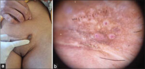
We report the case of a 35-year-old woman who presented with a painful lower abdominal multilobular nodule and skin discolorations around the abdominal incision site, two years after undergoing a cesarean section. The patient reported cyclic pain and volume augmentation of the nodule accompanied by red-colored fluid discharge from the incision site. Upon examination, a non-mobile, painful multilobular moderately pigmented nodule, measuring approximately 2×3 cm, was observed at the left lateral border of the incision (Fig, 1a). Dermoscopy revealed reddish areas separated by fibrous septa (Fig. 1b). Color Doppler ultrasound evaluation detected an irregular hypoechoic solid mass with internal vascularity, measuring 2×1.3×2.2 cm. Excision of the nodule confirmed the diagnosis of cutaneous endometriosis, and the patient was referred to gynecology for further evaluation of pelvic endometriosis.
 |
Figure 1: (a) Multilobular moderately pigmented nodule at the cesarean section scar. (b) Dermoscopy showing reddish areas separated by fibrous septa. |
Cutaneous endometriosis, a rare manifestation of endometriosis, is divided into primary and secondary forms, with the former resulting from spontaneous changes in specific tissues under unknown factors, and the latter caused by iatrogenic factors [1]. The incidence of secondary cutaneous endometriosis is about 3.5% in patients who undergo gynecological surgery and about 0.8% in women with a previous cesarean section [2]. Scar endometriosis, a type of secondary cutaneous endometriosis, is a rare condition that accounts for 0.03% to 0.15% of all cases of endometriosis in gynecological literature. Due to its varied presentation, such as pain, discoloration, and swelling around a Pfannenstiel skin incision, scar endometriosis often leads to a deferred diagnosis and unnecessary referrals. It can be mistaken for other conditions such as infections, abscesses, keloids, tumors, and lymphadenopathy [3].
This case highlights the challenges in diagnosing cutaneous endometriosis and emphasizes the importance of considering this condition in the differential diagnosis of abdominal wall lesions following cesarean sections. It also underscores the significance of timely excision as an effective treatment modality.
Consent
The examination of the patient was conducted according to the principles of the Declaration of Helsinki.
REFERENCES
1. Raffi L, Suresh R, McCalmont TH, Twigg AR. Cutaneous endometriosis. Int J Women Dermatol. 2019;5:384-6.
2. Nominato NS, Prates LF, Lauar I, Morais J, Maia L, Geber S. Caesarean section greatly increases risk of scar endometriosis. Eur J Obstet Gynecol Reprod Biol. 2010;152:83-5.
3. Gonzalez RH, Singh MS, Hamza SA. Cutaneous endometriosis:a case report and review of the literature. Am J Case Rep. 2021;22:e932493.
Notes
Request permissions
If you wish to reuse any or all of this article please use the e-mail (contact@odermatol.com) to contact with publisher.
| Related Articles | Search Authors in |
|
 http://orcid.org/0000-0003-3455-3810 http://orcid.org/0000-0003-3455-3810 http://orcid.org/0000-0002-5942-441X http://orcid.org/0000-0002-5942-441X |


Comments are closed.