A yellowish nodular rash under the dermoscope
Kenza Tahri Joutei Hassani , Zakia Douhi, Hanane Baybay, Sara Elloudi, Meryem Soughi, Fatima Zahra Mernissi
Department of Dermatology, University Hospital Hassan II, Fes, Morocco
Citation tools:


Copyright information
© Our Dermatology Online 2024. No commercial re-use. See rights and permissions. Published by Our Dermatology Online.
Primary localized cutaneous amyloidosis is the rarest form of cutaneous amyloidosis. Its prevalence is not well defined, with few cases reported in the literature. The mean age of onset is 55 years, without gender predilection [1]. Clinically, the lesions range from single to multiple nodules or infiltrated plaques that are smooth, shiny, waxy, orange yellow in appearance, sometimes hemorrhagic or bullous, and located on the lower limbs, face, scalp, or genitalia. On histological examination, the amyloid substance appears as a homogeneous amorphous and eosinophilic substance. Only Congo red staining can characterize amyloid deposits, with green birefringence under polarized light [2]. Immunohistochemistry allows to know the nature of the immunoglobulin light chains, Kappa, or Lambda. We found only two cases in the literature describing dermoscopy of ACNPL showing telangiectatic vessels on an orange-yellow background. The same dermoscopic patterns with elongated vessels on a yellow-orange background have been described in some cutaneous granulomatoses such as necrobiosis lipoidica, sarcoidosis and in histocytic disorders but not in ACNPL [3].
We describe a new case of dermoscopy of a primary localized cutaneous amyloidosis.
A 58-year-old woman, with a history of treated pulmonary tuberculosis and a 3-year history of asymptomatic lesions of the lower limbs and trunk without other associated signs. The examination found multiple yellow plaques, sometimes purplish or yellow violet, with a smooth surface, some of them anetodermal (Fig. 1a). Dermoscopy showed a homogeneous yellow pattern surrounded in places by telangiectatic vessels. Histopathologic study was compatible with ACNPL (Fig. 1b).
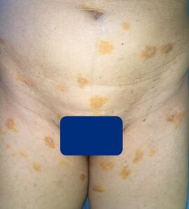 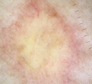 |
Figure 1: (a) Clinical picture showing multiple yellowish to yellow, purple plaques on the thigh and the trunk. (b) Dermoscopic picture showing: Homogeneous yellow pattern surrounded by telangiectatic vessels. |
Consent
The examination of the patient was conducted according to the principles of the Declaration of Helsinki.
REFERENCES
1. Schipper C, Cornelissen A, Welters C, Hoogbergen M. Treatment of rare nodular amyloidosis on the nose:a case report. JPRAS Open. 2015;6:25-30.
2. Kakani RS, Goldstein AE, Meisher I, Hoffman C. Nodular amyloidosis:case report and literature review. J Cutan Med Surg. 2001,5:101-4.
3. Bombonato C, Argenziano G, Lallas A, Moscarella E, Ragazzi M, Longo C. Orange color:a dermoscopic clue for the diagnosis of granulomatous skin diseases. J Am Acad Dermatol. 2015;72(1 Suppl):S60-3.
Notes
Request permissions
If you wish to reuse any or all of this article please use the e-mail (brzezoo77@yahoo.com) to contact with publisher.
| Related Articles | Search Authors in |
|
|
 http://orcid.org/0000-0003-3455-3810http://orcid.org/0000-0002-5942-441X http://orcid.org/0000-0003-3455-3810http://orcid.org/0000-0002-5942-441X |

Comments are closed.