Giant cutaneous herpes simplex with an atypical location in a woman with asthma receiving corticosteroids
Noura Kalmi , Zakia Douhi, Souad Choukri, Hanane Baybay, Sara Elloudi, Meryem Soughi, Fatima-Zahra Mernissi
Department of Dermatology, University Hospital Hassan II, Fes, Morocco
Citation tools:


Copyright information
© Our Dermatology Online 2024. No commercial re-use. See rights and permissions. Published by Our Dermatology Online.
A 28-year-old asthmatic patient on short-acting inhaled b2-agonists on request, with the notion of taking oral corticosteroids by self-medication at the time of the attacks. She was consulted for liquid lesions on the face that had been evolving for three days with a tingling sensation not preceded by the application of a topical. The patient had a history of herpes labialis.
The clinical examination found a patient in good general condition with a dermatological examination of a roughly oval plaque of 4.5 cm in size at the mid-frontal level with vesicles grouped in a cluster at the periphery and melicoid and hemorrhagic crusts in the center. The patient also had a slight bilateral conjunctival hyperemia (Fig. 1). The examination of the cervical lymph nodes revealed bilateral sub-mandibular adenopathies; the diagnosis of giant cutaneous herpes was retained. The patient was put on local fulcidic acid and oral valaciclovir at a dose of 1000 mg/dr for 5 days and eye drops by the ophthalmologists, with good improvement.
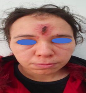 |
Figure 1: Clinical picture showing oval plaque of 4.5 cm in size at the mid-frontal level with vesicles grouped in a cluster at the periphery and melicoid and hemorrhagic crusts in the center. |
HSV is a common opportunistic infection in immunocompromised patients. Characteristically painful vesicles develop in a localized area like the lip, which subsequently go forward over days to form non-scarring scabs. The clinical expressions of HSV-1 infection in immunosuppressed hosts can be uncommon, including giant, erosive, ulcerative, vegetating, or hyperkeratotic lesions, with a higher incidence of chronicity, dissemination, recurrence, and acyclovir resistance [1,2]. It is explained by a number of host considerations such as age, immune status, intercurrent disease, and the presence of a preceding skin illness [3].
Intravenous antiviral therapy may be required in immunocompromised patients and those with severe disseminated infections [1]. However, in our case, the oral treatment was sufficient with a satisfactory response.
Consent
The examination of the patient was conducted according to the principles of the Declaration of Helsinki.
REFERENCES
1. Khera P, Haught JM, McSorley J, English JC 3rd. Atypical presentations of herpesvirus infections in patients with chronic lymphocytic leukemia. J Am Acad Dermatol. 2009;60:484-6.
2. Metin A, Kanik-Yuksek S, Ozkaya-Parlakay A, Tezer H. Giant Herpes Labialis in a Child with DOCK8-deficient Hyper-IgE Syndrome. Pediatr Neonatol. 2016;57:79-80.
3. Qian YT, Ma JY, Liu JW, Liu W, Ma DL. Chronic Herpes Simplex virus infection in an immunosuppressed girl. J Pediatr. 2019;212:237-237.e1.
Notes
Request permissions
If you wish to reuse any or all of this article please use the e-mail (brzezoo77@yahoo.com) to contact with publisher.
| Related Articles | Search Authors in |
|
|
 http://orcid.org/0000-0003-3455-3810http://orcid.org/0000-0002-5942-441X http://orcid.org/0000-0003-3455-3810http://orcid.org/0000-0002-5942-441X |

Comments are closed.