Molluscum contagiosum scrotale in a child
Imane Couissi , Hanane BayBay, Zineb Fajri, Meryem Soughi, Zakia Douhi, Sara El Loudi, Fatima Zahra Mernissi
Department of Dermatology, University Hassan II, Fès, Morocco
Citation tools:


Copyright information
© Our Dermatology Online 2024. No commercial re-use. See rights and permissions. Published by Our Dermatology Online.
Molluscum contagiosum (MC) is a highly contagious benign viral epidermal infection caused by a DNA virus of the Poxviridae family, Molluscipox genus.
It usually affects school-aged children and occasionally adults and immunocompromised individuals [1].
Clinically, it appears as papules or nodules with umbilicated centers 1 to 10 mm in diameter, don’t exceed 1 cm. However, an atypical presentation in terms of location, size, and number can be found and increases the difficulty of diagnosis.
In children, MC is often located in the face, trunk, and extremities. However, localization in the genital area is more common in adults and is considered a sexually transmitted disease [2].
Therefore, cases of genital MC must rule out child abuse, even though self-inoculation of poxvirus is a potential mode of transmission.
We report a case of a 9-year-old child who presented for 6 months with well-limited rounded nodules and papules umbilicated in places with smooth surfaces at the scrotal and inguinal level (Fig. 1a), with no central polylobular white-yellow structureless area at the dermoscopy whose histopathological findings were in favor of a molluscum contagiosum (Fig. 1b).
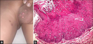 |
Figure 1: (a) Clinical image showing well-limited rounded nodules and papules umbilicated in places with a smooth surface at the scrotal and inguinal level. (b) Histological image, HES G staining × 100 showing a piriform epidermal invagination with intrakeratinocyte globoid inclusions: molluscoid corpuscles becoming basophilic and stacked then rejected on the surface. |
The transmission of molluscum contagiosum is caused by direct physical contact.
Swimming, co-bathing, fomite by sharing towels/sponges, and self-inoculation, promote MC spread. Weismann et al reported that sitting around the pool is a possible mode of transmission [3].
Consent
The examination of the patient was conducted according to the principles of the Declaration of Helsinki.
The authors certify that they have obtained all appropriate patient consent forms, in which the patients gave their consent for images and other clinical information to be included in the journal. The patients understand that their names and initials will not be published and due effort will be made to conceal their identity, but that anonymity cannot be guaranteed.
REFERENCES
1. Chen X, Anstey AV and Bugert JJ. Molluscum contagiosum virus infection. Lancet Infect Dis. 2013;13:877–88.
2. Ishikawa MK, Arps DP, Chow C, Hocker TL, Fullen DR. Histopathological features of molluscum contagiosum other than molluscum bodies. Histopathology. 2015;67:836–42.
3. Vora RV, Pilani AP and Kota RK. Extensive giant Molluscum contagiosum in a HIV positive patient. J Clin Diagn Res. 2015;9:Wd01–2.
Notes
Request permissions
If you wish to reuse any or all of this article please use the e-mail (brzezoo77@yahoo.com) to contact with publisher.
| Related Articles | Search Authors in |
|
|
 http://orcid.org/0009-0005-2261-6919http://orcid.org/0000-0003-3455-3810http://orcid.org/0000-0002-5942-441X http://orcid.org/0009-0005-2261-6919http://orcid.org/0000-0003-3455-3810http://orcid.org/0000-0002-5942-441X |

Comments are closed.