Mucosal involvement in the context of mycosis fungoides revealing a cicatricial pemphigoid
Imane Couissi , Sara El Loudi, Kawtar El Fid, Meryem Soughi, Zakia Douhi, Hanane BayBay, Fatima Zahra Mernissi
Department of Dermatology, University Hospital Hassan II, Fès, Morocco
Citation tools:


Copyright information
© Our Dermatology Online 2024. No commercial re-use. See rights and permissions. Published by Our Dermatology Online.
Sir,
Cicatricial pemphigoid (CP), also known as cicatricial pemphigoid, is a rare, synechial, subepidermal autoimmune bullous dermatosis characterized clinically by its elective involvement of mucous membranes, most often the oral and ocular mucosae. Because of the risk of serious complications, such as blindness and airway involvement, early and aggressive treatment may be warranted. Recent studies have shown the association of CP with an increased risk of solid cancer.
Herein, we report a sixty-year-old female who had been followed at our department for four years for a mycosis fungoid at tumor stage transformed classified as T3N0M0 or stage IIB. The patient received nine courses of gemcitabine followed by localized radiotherapy on two persistent nodules on the left thigh with complete remission. Sixteen months later, the patient developed oral erosions, epistaxis with high dysphagia, and bilateral ocular redness.
Clinical examination found two well-limited, erythematous ulcerations on the soft palate in the process of healing (Fig. 1a), conjunctival hyperemia with bilateral pterygium (Fig. 1b), and an inflammatory nasal mucosa with a well-limited, rounded erosion on the right nostril mucosa (Fig. 1c).
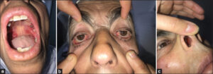 |
Figure 1: (a) Two well-limited, erythematous ulcerations on the soft palate in the process of healing.(b) Conjunctival hyperemia with bilateral pterygium.(c) An inflammatory nasal mucosa with a well-limited, rounded erosion on the right nostril mucosa. |
An ophthalmological examination revealed fibrotic conjunctivitis evoking cicatricial pemphigoid at first. Laryngoscopy revealed whitish ulcerations at the base of the tongue and on the laryngeal surface of the epiglottis. A biopsy with a histological study and direct immunofluorescence of an oral bulla was compatible with cicatricial pemphigoid.
The patient was put on corticosteroid bolus and then low-dose corticosteroid therapy associated with a course of rituximab. The evolution was marked by an improvement in oral, ocular, and nasal involvement.
Cicatricial pemphigoid (CP) is an autoimmune disease characterized by the presence of various autoantibodies directed against basement membrane antigens, most frequently BP180 yet also BP230 or b4 integrin, with a minority against laminin 5 [1].
In the literature, several retrospective studies have reported an association between CP and malignancy, especially solid tumors.
Egan et al. described 35 patients with CP followed over a twelve-year period; ten patients (28.6%) developed solitary cancer. Eight of these patients developed cancer after the onset of CP, most within 12 to 14 months, and all deaths were cancer-related [2].
Associated malignancies included three lung, three stomach, two colon, and two endometrial cancers.
In addition to solid cancers, CP was described by Sadler et al. in a patient with mycosis fungoides treated for twelve years with topical clobetasol, who developed oral and nasal erosions. Histopathology confirmed the diagnosis of CP. This was the first association between CP and lymphoma [3].
This association was later documented in another patient, with diffuse large B-cell non-Hodgkin’s lymphoma in a series by Shannon et al. [4].
Our case was the third reported association between CP and lymphoma.
Most studies found the association between CP with laminin-5 and neoplasia.
Some reports suggested that CP may represent a paraneoplastic syndrome.
Various pathogenetic hypotheses have been put forward, including the theory that tumor cells secrete anti-laminin-5 antibodies directed against the skin and cause bulla formation [5].
In our case, the patient presented CP sixteen months after the diagnosis of mycosis fungoides with oral, ocular, nasal, and laryngeal involvement, which may be considered a paraneoplastic syndrome.
Various studies have demonstrated the correlation between laminin-5 in CP and cancer, emphasizing the importance of the immunological diagnosis of these autoantibodies.
However, the absence of this antigen in other cases casts doubt on this hypothesis, hence the need for further studies.
Consent
The examination of the patient was conducted according to the principles of the Declaration of Helsinki.
The authors certify that they have obtained all appropriate patient consent forms, in which the patients gave their consent for images and other clinical information to be included in the journal. The patients understand that their names and initials will not be published and due effort will be made to conceal their identity, but that anonymity cannot be guaranteed.
REFERENCES
1. Kartan S, Shi VY, Clark AK, Chan LS. Paraneoplastic pemphigus and autoimmune blistering diseases associated with neoplasm:Characteristics, diagnosis, associated neoplasms, proposed pathogenesis, treatment. Am J Clin Dermatol. 2017;18:105-26.
2. Amber KT, Bloom R, Hertl M. A systematic review with pooled analysis of clinical presentation and immunodiagnostic testing in mucous membrane pemphigoid:Association of anti-laminin-332 IgG with oropharyngeal involvement and the usefulness of ELISA. J Eur Acad Dermatol Venereol. 2016;30:72-7.
3. Balestri R, Magnano M, La Placa M. Malignancies in bullous pemphigoid:A controversial association. J Dermatol. 2016;43:125-33.
4. Ding DC, Chu TY, Hsu YH. Remission of anti-epiligrin cicatricial pemphigoid after excision of cervical adenocarcinoma. J Cutan Pathol. 2014;41:692-3.
5. Taylor J, McMillan R, Shephard M. World workshop on oral medicine VI:A systematic review of the treatment of mucous membrane pemphigoid. Oral Surg Oral Med Oral Pathol Oral Radiol. 2015;120:161-71.
Notes
Request permissions
If you wish to reuse any or all of this article please use the e-mail (contact@odermatol.com) to contact with publisher.
| Related Articles | Search Authors in |
|
|
 http://orcid.org/0009-0005-2261-6919http://orcid.org/0000-0002-5942-441Xhttp://orcid.org/0000-0003-3455-3810 http://orcid.org/0009-0005-2261-6919http://orcid.org/0000-0002-5942-441Xhttp://orcid.org/0000-0003-3455-3810 |

Comments are closed.