Adult tinea in a psoriatic patient
Soukaina Karimi, Maryem Aboudourib, Ouafa Hocar, Said Amal
Dermatology departement, Arrazi Mohammed VI universal Hospital, Marrakech, Morocco
 , MD, E-mail: Soukaina.karimi@gmail.com
Citation tools:


Copyright information
© Our Dermatology Online 2024. No commercial re-use. See rights and permissions. Published by Our Dermatology Online.
Sir,
Tinea is a superficial fungal infection of the hair follicles caused by the filamentous fungi dermatophytes that have distinct predilection parasitism for the hair. This infection affects predominantly preadolescent children and is rarely seen in adults. Herein, we present a case of tinea capitis in an adult.
A 64-year-old, menopaused female patient had psoriasis and vitiligo for more than twenty years and was treated with local steroids and phototherapy with a good clinical response. She presented with pruritic erythematous and squamous lesions on the face apparent for two months. A clinical examination revealed multiple, rounded, erythematous, and slightly scaly plaques on the periphery, located on the forehead, eyebrows, tip of the nose, and both cheeks (Fig. 1a). On the scalp, there were three partially alopecic, erythematous plaques surmounted by fine, yellowish scales located in the frontal and temporal area, among which the largest measured 4 cm/3 cm (Figs. 1b and 1c).
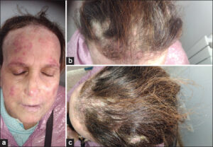 |
Figure 1: (a)Clinical examination of the face: multiple erythematous and scaly plaques of small sizes, localized on the forehead, eyebrows, tip of the nose, and the cheeks. (b) Clinical examination of the scalp: three alopecic, erythematous plaques with yellowish scales, localized on the temporal and parietal areas, multiple broken hairs seen, among which the largest measured 4 cm/3 cm. (c) Clinical examination of the scalp: a top view of the alopecic, erythematous plaques with yellowish scales in the frontal and temporal areas. |
Trichoscopy showed an erythematous background, yellow scales, and corkscrew hair (Fig. 2). The diagnosis of a fungal infection was strongly suspected.
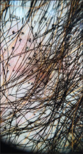 |
Figure 2: Trichoscopic image (DermLite 4; 3Gen; polarized mode, 10×): erythematous background, yellow scales, and multiple corkscrew hairs. |
Direct microscopic examination of the face and hair scales revealed numerous spores with endothrix parasitism, and a cultural examination isolated Trichophyton violaceum after three weeks.
On the basis of the clinico-dermoscopic and microbiological results, the diagnosis of mycosis of the scalp and skin was established.
Therapeutic management consisted of griseofulvin at 1 g per day for six weeks. A follow-up noted the total disappearance of the facial lesions, the regression of the dermoscopic signs, and the beginning of hair regrowth (Figs. 3a and 3b).
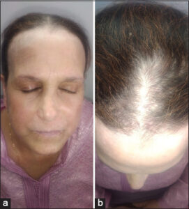 |
Figure 3: (a) Follow-up clinical examination of the face after six weeks: regression of the facial lesions and pruritus. (b) Follow-up clinical examination of the scalp after six weeks: regression of the pruritus, erythema, and beginning of hair regrowth. |
Tinea capitis is a common childhood fungal infection. The exact incidence in adults is unknown yet is generally thought to be low. Only 3% to 5% of cases of tinea capitis occur after puberty [1].
Its clinical manifestations range from mild scaling with little hair loss to large inflammatory and pustular plaques with extensive alopecia [2]. Moreover, it is often misdiagnosed in adults because of its atypical clinical presentation [3].
Trichoscopy (hair dermoscopy) is highly useful in detecting ringworm of the scalp, with a sensitivity of 94% and a specificity of 83%. Its characteristic features include comma hairs, corkscrew hairs, zigzag hairs, Morse code-like hairs, bent hairs, block hairs, I-hairs, and whitish sheaths [4].
In adults, Trichophyton tonsurans is the primary fungal species responsible for tinea capitis in the U.S., Canada, Mexico, and Central America. Meanwhile, Trichophyton violaceum is more prevalent in Africa, India, Thailand [5], and Iran [6], which was similar to our patient. In some European and Asian countries, including Korea, Microsporum canis is the main pathogen identified [5].
Multiple risk factors have been described in the series on tinea in adults: diabetes, immunosuppressants, prolonged use of tropical or systemic steroids, and anemia [6]. The other predisposing factors were contact with animals or direct contact with an infected person [7].
In numerous reports, a higher incidence of tinea in adults (46.4%) was given in postmenopausal females, and this is explained by the involution of the sebaceous glands following a decrease in the levels of blood estrogen [7].
Regarding our patient, two underlying factors were found: 1) the application of topical steroids one month before the appearance of the lesions 2) and menopause, which explains this occurrence.
The treatment of tinea capitis relies on the use of terbinafine, itraconazole, griseofulvin, and fluconazole. It is usually effective yet should be prescribed for at least one month [5].
Adult tinea corporis has an unusual clinical and mycological presentation. Trichoscopy is helpful in increasing the sensitivity of physical examination. Thus, an early diagnosis and treatment is essential to avoid permanent damage to the hair follicle and, thus, prevent scarring alopecia.
Consent
The examination of the patient was conducted according to the principles of the Declaration of Helsinki.
The authors certify that they have obtained all appropriate patient consent forms, in which the patients gave their consent for images and other clinical information to be included in the journal. The patients understand that their names and initials will not be published and due effort will be made to conceal their identity, but that anonymity cannot be guaranteed.
REFERENCES
1. Martin ES, Elewski BE. Tinea capitis in adult women masquerading as bacterial pyoderma. J Am Acad Dermatol. 2003;49:177 9.
2. Panigrahi A, Sil A, Biswas SK. Tinea capitis:Bedside diagnosis by dermoscopy. J Pediatr. 2020;222:248.
3. Torres Guerrero E, Espinoza Hernández CJ, Arroyo Camarena S, Atoche Diéguez CE. Tinea caused by Microsporum gypseum. Our Dermatol Online. 2018;9:380-5.
4. Lim SS, Shin K, Mun JH. Dermoscopy for cutaneous fungal infections:A brief review. Health Sci Rep. 2022;5:e464.
5. Park S, Park S, Yun S, Kim H, Park J. Tinea capitis in adults:A 18-year retrospective, single-centre study in Korea. Mycoses. 2019;62:609 16.
6. Khosravi AR, Shokri H, Vahedi G. Factors in etiology and predisposition of adult tinea capitis and review of published literature. Mycopathologia. 2016;181:371 8.
7. El-Khalawany M, Shaaban D, Hassan H, Abdalsalam F, Eassa B, Abdel Kader A, et al. A multicenter clinicomycological study evaluating the spectrum of adult tinea capitis in Egypt. Acta Dermatovenerol Alp Pannonica Adriat. 2013;22:77-82.
Notes
Request permissions
If you wish to reuse any or all of this article please use the e-mail (brzezoo77@yahoo.com) to contact with publisher.
| Related Articles | Search Authors in |
|
|
|

Comments are closed.