Norwegian scabies in a patient with Down syndrome: A case report
Alina Werner, Joanna Zalewska , Anna Płatkowska, Elwira Paluchowska, Witold Owczarek
Dermatology Clinic, Military Institute of Medicine – National Research Institute, Warsaw, Poland
Citation tools:


Copyright information
© Our Dermatology Online 2024. No commercial re-use. See rights and permissions. Published by Our Dermatology Online.
ABSTRACT
Scabies is a highly infectious dermatosis. It is caused by the mite Sarcoptes scabiei var. hominis. The available estimates indicate more than 400 million cases of scabies annually. The main route of infection is direct skin contact. The main symptoms such as pruritus appear 3–6 weeks after infection. Additionally, papules, erythema, erosions, excoriations, and crusts may be visible. One of the rarest forms of scabies is Norwegian scabies. This is the most severe variant. Its clinical picture may resemble psoriasis. In the article, we present the case of a 21-year-old patient with Down syndrome who was admitted to the department of dermatology with a diagnosis of psoriasis. During hospitalization, due to clinical doubts, it was decided to extend the diagnostics. Basing on the microscopic examination, Norwegian scabies was diagnosed. The treatment involved oral ivermectin and topical 5% permethrin, which resulted in a remission of the skin lesions and the resolution of itching.
Key words: Sarcoptes scabiei, Scabies, Crusted scabies, Permethrin, Ivermectin
INTRODUCTION
Scabies is a widespread, infectious disease. It is caused by the mite Sarcoptes scabiei var. hominis. The available estimates indicate more than 400 million cases of scabies annually [1]. Scabies occurs in all latitudes, so all populations are at risk of developing the disease. The risk is much higher among people living in crowded and pauper conditions, due to greater skin-to-skin contact and lack of access to effective treatment. The highest prevalence is observed among infants and children in tropical countries [2].
Infestation usually occurs with 5–15 specimens. The entire lifecycle takes place in the human epidermis. Fertilized females burrow into the epidermis where they lay their eggs. One female lays about 1–20 eggs and dies after several weeks. The larvae hatch after about sixty hours, and their development into an adult form takes about two weeks. The main route of infection is direct skin contact. Symptoms appear 3–6 weeks after infection. Hence, to locate its source, while collecting medical history, a longer period should be investigated. In the case of reinfestation, symptoms appear much faster [3]. The main symptom occurs as pruritus, which intensifies after warming the body, especially at night. Additionally, papules, erythema, erosions, excoriations, and scabs may be visible.
They result from the presence of the parasite itself, as well as from type IV hypersensitivity reactions. The lesions are most often located around the interdigital areas, torso, and buttocks. Moreover, it is worth adding that zoonotic infections (referred to as pseudoscabies) are rare and self-limiting. It is commonly thought that scabies mites inhabiting other species cannot complete their full lifecycle in the human epidermis. However, experimental human infections with scabies mites of dog origin resulted in a successful replication with hatching and the development of mites. Additionally, there are several case reports mentioning spontaneous zoonotic scabies [4].
One of the rarest forms of scabies is Norwegian scabies, also known as hyperkeratotic scabies. This is the most severe variant and is highly infectious. Symptoms include the presence of a layered, thickened scale. The clinical picture may resemble psoriasis [5–8]. The treatment of Norwegian scabies includes combined oral and topical antiparasitic agents [9].
CASE REPORT
A 21-year-old patient with Down syndrome was admitted to the Dermatology Clinic of the Military Institute of Medicine with a diagnosis of psoriasis in December 2021.
A medical history was collected from his mother. The patient had suffered from psoriasis since he was twelve years old (2013), the diagnosis was confirmed by histopathological examination. The previous treatment included topical corticosteroids and oral acitretin (25 mg a day) from April to December 2019. At that time, due to a difficult family situation, the patient was temporarily staying at an orphanage, so a reliable treatment effectiveness assessment was difficult to determine. Moreover, the reason for its discontinuation was unknown. Since 2020, the patient had again been under his mother’s care. He did not receive systemic treatment, only topical one with corticosteroids. The skin lesions worsened approximately one month before hospitalization.
The clinical picture was dominated by massive, hyperkeratotic layers with fissures and moderate inflammation of the skin (Figs. 1a – 1c). Moreover, lesions covered with thick layers of scale were found on the scalp and chest (Fig. 2). Numerous dermal and epidermal papules and erythema were visible on the genitals and groins. The skin lesions were accompanied by intense itching, which was reflected in numerous excoriations. All fingernails and toenails were affected: massive subungual hyperkeratosis was observed (Fig. 3).
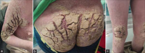 |
Figure 1: (a) Fissures around the right shoulder. (b) Fissures at the sacrolumbar area. (c) Fissures around the knees. |
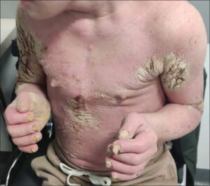 |
Figure 2: Thick scale on the chest. |
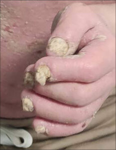 |
Figure 3: Subungual hyperkeratosis. |
The patient’s mother presented discreet skin lesions, such as papules and excoriations, on the trunk.
During hospitalization, scrapings were taken from the superficial layers of the hyperkeratotic lesions. Microscopic evaluation confirmed the presence of Sarcoptes scabiei. Norwegian scabies was the diagnosis. Performed laboratory tests revealed eosinophilia (4.37 x 10³/μL) and increased total IgE concentration (338 IU/mL). Additionally, hepatitis B and C and HIV infections were excluded. The patient’s mother underwent a dermatoscopic examination, which revealed burrows. The woman was diagnosed with the classic form of scabies.
The patient was treated orally with ivermectin at a single dose of 200 ug/kg of body weight and topically with 5% permethrin at a single dose of 30 g on the entire skin surface, including the scalp and face. Additionally, local keratolytic medications were implemented, which resulted in a significant reduction of hyperkeratotic build-ups (Figs. 4a – 4e) and resolution of erythematous lesions and itching. The mother was treated topically with permethrin, excluding the scalp and face.
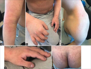 |
Figure 4: (a) Right arm after treatment. (b) Right hand after treatment. (c) Left arm after treatment. (d) Left hand and nails after treatment. (e) Sacrolumbar area after treatment. |
The patient was discharged from the clinic with a recommendation to repeat treatment with ivermectin and permethrin eight days after the first dose and to attend for a follow-up visit in four weeks. The patient and his mother showed up to the outpatient clinic five months after hospitalization, without any symptoms. Due to the clinical improvement, a follow-up microscopic examination was waived, and both were considered cured.
DISCUSSION
Rare forms of scabies include the Norwegian, nodular, and bullous variants. Norwegian scabies, also called crusted scabies, is the most severe one. It affects patients with immune deficiencies, after transplantations, patients with Down syndrome, HIV-infected, as well as patients who chronically use topical corticosteroids. This variant is highly infectious and is associated with mortality [10]. A disturbed and insufficient immune response allows the parasite to multiply unrestricted. Infection may also easily occur indirectly, through exfoliated epidermis. The clinical picture is dominated by thick, layered, extensive scales or crusts. Norwegian scabies may cause erythroderma, affecting the scalp, ears, palms, and soles of the feet. In addition, mites may also accumulate in the matrix of the nails, causing onychodystrophy, subungual hyperkeratosis, or changes in the color of the nail plates. These symptoms may be confused with other diseases such as psoriasis or nail fungus [11], while contributing to prolonged infestations [12].
The diagnostics of scabies involves the microscopic examination of scrapings from the superficial layers of the skin. It allows to detect the presence of mites, eggs, or brown feces. It is recommended to take several samples from characteristic skin lesions with a scalpel, preferably using oil, which helps the material to adhere to the blade [13]. In the classic form of scabies, there are about 10–20 parasites in the patient’s skin. A negative test result does not exclude the diagnosis. However, in the case of Norwegian scabies, massive infestations occur, and the number of parasites reaches approx. 1–2 million, thus the probability of a positive test is much higher. Other diagnostic methods, such as the burrow ink test have been described in the literature [14]. It is a simple and often overlooked test in which the ink is gently rubbed into the suspected area. Its excess is wiped off with an alcohol-soaked cotton swab and the burrow becomes visible as a wavy line [15]. Other methods include dermatoscopy and videodermatoscopy. A new approach to scabies diagnosis is serological testing. In 2011, Rama et al. developed a specific IgE antibody against a recombinant scabies antigen. In this study, the sensitivity and specificity were 100% and 93.75%, respectively [16]. Laboratory tests may also be helpful. An increase in total IgE concentration and peripheral eosinophilia are distinctive [17].
Norwegian scabies due to its non-specific presentation and, consequently, delayed diagnosis may cause numerous complications such as secondary bacterial infections, cellulitis, or sepsis, which is associated with high mortality [18,19]. Moreover, invasive Staphylococcus aureus and Streptococcus pyogenes infections may result in kidney disease and rheumatic heart disease [2].
The 2017 European guidelines for the management of Norwegian scabies recommend 5% permethrin or 25% benzyl benzoate repeated daily for seven days, then twice a week until cure in combination with oral ivermectin 200 μg/kg on days 1, 2, and 8. For severe cases, when a follow-up microscopic examination gives positive results, additional ivermectin treatment might be required on days 9 and 15 or on days 9, 15, 22, and 29 [9].
In Poland, according to the summary of product characteristics, a single oral dose of ivermectin is 200 μg/kg of body weight, and in the case of Norwegian scabies, a second dose within 8 to 15 days and/or simultaneous local treatment. The recovery occurs four weeks after treatment. Persistent itching does not justify repeating the treatment before this date. The administration of a second dose should be considered within two weeks of the administration of the first dose if new specific lesions appear or parasitological examination gives a positive result.
All people living in the same household should be treated, regardless of the presence of clinical symptoms. Patients with Norwegian scabies should be isolated until the end of treatment [20]. The infestation is considered cleared if one week after the end of treatment there are no skin lesions or nocturnal itching. However, pruritus may persist up to 2-4 weeks after treatment. It should be cured with the repeated application of emollients. Oral antihistamines and mild topical corticosteroids may also be helpful. A follow-up visit is recommended two weeks after the end of treatment to perform a control microscopic examination [9].
An inseparable part of scabies treatment includes hygiene recommendations such as clothes, bedding, and towels washing at 60°C at least. Everyday objects that cannot be washed should be placed in plastic bags for at least seven days. All smooth surfaces should be washed thoroughly, and upholstered furniture ought to be vacuumed. Mattresses may be insulated with painting foil. The ability of scabies mites to survive in beds and upholstery (away from the human host) is up to four days [3].
The differential diagnosis of Norwegian scabies should include psoriasis, atopic dermatitis, chronic allergic eczema, seborrheic dermatitis, lichen planus, palmoplantar keratosis, and cutaneous lymphoma [21]. The correlation between Down syndrome and psoriasis is vague. The prevalence of psoriasis in patients with Down syndrome ranges from 0.5% to 8% [22], and in turn from 2% to 3% in the world population [23]. Nevertheless, it should be noted that patients with trisomy 21 have a higher risk of developing skin diseases [24].
CONCLUSION
In this article, we present a case of Norwegian scabies, which posed diagnostic difficulties due to its occurrence in a patient suffering from psoriasis. The clinical picture of both diseases may be similar. Moreover, Norwegian scabies is a rare disease yet cannot be omitted in the differential diagnosis. Appropriate treatment not only helps to relieve symptoms yet also limits further transmission of the infection. The presented case confirmed the effectiveness of combined therapy with ivermectin and permethrin in the treatment of Norwegian scabies.
Consent
The examination of the patient was conducted according to the principles of the Declaration of Helsinki.
The authors certify that they have obtained all appropriate patient consent forms, in which the patients gave their consent for images and other clinical information to be included in the journal. The patients understand that their names and initials will not be published, and due effort will be made to conceal their identity, but that anonymity cannot be guaranteed.
REFERENCES
1. Karimkhani C, Colombara DV, Drucker AM, Norton SA, Hay R, Engelman D, et al. The global burden of scabies:A cross-sectional analysis from the Global Burden of Disease Study 2015. Lancet Infect Dis. 2017;17:1247-54.
2. WHO informal consultation on a framework for scabies control, World Health Organization Regional Office for the Western Pacific, Manila, Philippines, 19–21 February 2019:meeting report.
3. Banerji A. Scabies. Paediatrics Child Health. 2015;20:395-402.
4. Moroni B, Rossi L, Bernigaud C, Guillot J. Zoonotic episodes of scabies:A global overview. Pathogens. 2022;11:213.
5. Kosmala A, Żaba R, Adamski Z. Scabies:The great imitator?Recent reports on scabies and a case report. Dermatol Rev. 2017;9:43-51.
6. Fonseca V, Price HN, Jeffries M, Alder SL, Hansen RC. Crusted scabies misdiagnosed as erythrodermic psoriasis in a 3-year-old girl with Down syndrome. Pediatr Dermatol. 2014;31:753-4.
7. Goyal NN, Wong GA. Psoriasis or crusted scabies. Clin Exp Dermatol. 2008;33:211-2.
8. Gach JE, Heagerty A. Crusted scabies looking like psoriasis. Lancet. 2000;356:650.
9. Salavastru CM, Chosidow O, Boffa MJ, Janier M, Tiplica GS. European guideline for the management of scabies. J Eur Acad Dermatol Venereol. 2017;31:1248-53.
10. Lynar S, Currie BJ, Baird R. Scabies and mortality. Lancet Infect Dis. 2017;17:1234.
11. Tempark T, Lekwuttikarn R, Chatproedprai S, Wananukul S. Nail scabies:An unusual presentation often overlooked and mistreated. J Trop Pediatr. 2017;63:155-9.
12. Abdullah L, Abbas O. Common nail changes and disorders in older people:Diagnosis and management. Can Fam Physician. 2011;57:173-81.
13. Shimose L, Munoz-Price LS. Diagnosis, prevention, and treatment of scabies. Curr Infect Dis Rep. 2013;15:426-31.
14. Rauwerdink D, Balak D. Burrow ink test for scabies. N Engl J Med. 2023;7;389.
15. Leung V, Miller M. Detection of scabies:A systematic review of diagnostic methods. Can J Infect Dis Med Microbiol. 2011;22:143-6.
16. Jayaraj R, Hales B, Viberg L, Pizzuto S, Holt D, Rolland JM, et al. A diagnostic test for scabies:IgE specificity for a recombinant allergen of Sarcoptes scabiei. Diagn Microbiol Infect Dis. 2011;71:403-7.
17. Roberts LJ, Huff am SE, Walton SF, Currie BJ. Crusted scabies:Clinical and immunological findings in seventy-eight patients and a review of the literature. J Infect. 2005;50:375-81.
18. Engelman D, Kiang K, Chosidow O, McCarthy J, Fuller C, Lammie P, et al. Toward the global control of human scabies:Introducing the International Alliance for the Control of Scabies. PLoS Negl Trop Dis. 2013;7:e2167.
19. Apap C, Piscopo T, Boffa MJ. Crusted (Norwegian) scabies treated with oral ivermectin:A case report and overview. Malta Med J. 2021;25:49-53.
20. Kosmala A, Szymoniak-Lipska M, Jałowska M, Dobrzyńska M, Bowszyc-Dmochowska M, Adamski Z, et al. Crusted scabies in a patient with systemic disorders:Evaluation of ivermectin treatment results. Dermatol Rev. 2019;106:671-9.
21. Matsuura H, Senoo A, Saito M, Fujimoto Y. Norwegian scabies. Cleve Clin J Med. 2019;86:163-4.
22. Madani A, Almuhaideb Q. Adalimumab therapy in a patient with psoriasis, Down syndrome, and concomitant Hepatitis B Virus infection. Biologics. 2021;15:375-8.
23. Damiani G, Bragazzi NL, Karimkhani Aksut C, Wu D, Alicandro G, McGonagle D, et al. The global, regional, and national burden of psoriasis:Results and insights from the Global Burden of Disease 2019 Study. Front Med (Lausanne). 2021;8:743180.
24. Lam M, Lu JD, Elhadad L, Sibbald C, Alhusayen R. Common dermatologic disorders in Down syndrome:Systematic review. JMIR Dermatol. 2022;5:e33391.
Notes

Comments are closed.