Cutaneous schwannoma with atypical location
Sara Kerroum , Zineb Zeggwagh, Nadia Ismaili, Mariame Meziane, Laila Benzekri, Karima Senouci
Department of Dermatology, Mohammed V University in Rabat, Ibn Sina University Hospital, Rabat, Morocco
Citation tools:


Copyright information
© Our Dermatology Online 2023. No commercial re-use. See rights and permissions. Published by Our Dermatology Online.
Schwannomas are benign, slow-growing nerve tumours that develop from schwann cells. Schwann cells play an important role in the propagation of nerve impulses by encapsulating the nerve fibres of peripheral nerves, cranial nerves and nerves of the autonomic system [1]. Head and neck schwannomas account for 25-45% of all schwannomas and are dominated by vestibular schwannomas. Clinically, cutaneous schwannomas are generally asymptomatic, presenting as a slowly growing mass causing cosmetic damage; neurological signs such as pain or paresthesia may be found in one third of cases.Histologically, schwannomas are characterised by a proliferation of elongated spindle cells, with elongated nuclei often arranged in a palisade pattern called Antoni A type (Verocay corps), while those in which the cells are loose and irregularly arranged are called Antoni B type [2]. Immunohistochemical study often shows positive staining for S-100 protein [3]. The treatment of choice is surgical removal. Dissection of this type of tumour is facilitated by the presence of a capsule which forms a smooth surface under the skin. Local recurrence has rarely been reported.
A 30-year-old patient with no notable pathological history who has had a rounded (Fig. 1a), pearly white asymptomatic nodule above the upper lip for about 3 months (Fig. 1b). The patient consulted for major cosmetic damage. An excisional biopsy was performed showing a histological and immunohistochemical appearance in favour of a cutaneous schwannoma. Annual surveillance was recommended.
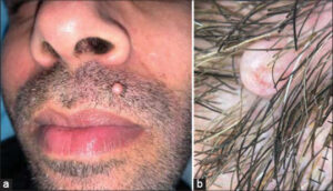 |
Figure 1: (a) Regular rounded nodule above the upper lip. (b) Pearly white dermoscopic appearance. |
Consent
The examination of the patient was conducted according to theprinciples of the Declaration of Helsinki.The authors certify that they have obtained all appropriate patientconsent forms, in which the patients gave their consent for imagesand other clinical information to be included in the journal. Thepatients understand that their names and initials will not bepublished and due effort will be made to conceal their identity,but that anonymity cannot be guaranteed.
REFERENCES
1. Benchafai I, Fetouhi A, Allaoui M, Ouraini S, Jahidi A, Benariba F:Schwannoma of the ear lobe:an etiology that should be taken into consideration in patients. Case report. Pan Frican Med J. 2020;2:1.
2. Cotran RS, Kumar V. Robbins and Cotran Pathologic Basis of Disease. 7th ed. Philadelphia:Saunders Elsevier;2005. Peripheral nerve sheath tumors;pp. 1411–3.
3. Tzu-Chun L, Po-Yuan W, Tze-Yi L, Tsong-Liang L. An infrequent plexiform variant of schwannoma of the glans penis:a rare finding. Asian J Androl. 2010;12:455–7.
Notes
Request permissions
If you wish to reuse any or all of this article please use the e-mail (brzezoo77@yahoo.com) to contact with publisher.
| Related Articles | Search Authors in |
|
|
|

Comments are closed.