Clinical Images of a Morbihan disease associated to scrotal lymphedema
Nada Bennouna , Fatimazahra Elfetouaki, Fouzia Hali, Soumiya Chiheb
Department of Dermatology and Venerology, Ibn Rochd University Hospital Casablanca, Morocco
How to cite this article: Bennouna N, Elfetouaki F, Hali F, Chiheb S. Clinical Images of a Morbihan disease associated to scrotal lymphedema. Our Dermatol Online. 2023;14(e):e8.
Submission: 01.11.2022; Acceptance: 05.05.2023
DOI: 10.7241/ourd.2023e.8
Citation tools:


Copyright information
© Our Dermatology Online 2023. No commercial re-use. See rights and permissions. Published by Our Dermatology Online.
Morbihan disease, also known as rosaceous lymphedema, is characterized by persistent erythema and solid edema of the upper two thirds of the face. Genital elephantiasis is characterized by massive growth of genitals [1] as a result of chronic lymphedema caused by numerous infectious and noninfectiuos triggers [2].
We report a case of a 52-year-old man presented with a 2-year history of swelling and redness on the upper part of his face. He also had a 6-year history of rosacea and rhinophyma without any treatment. He was recently diagnosed with primary scrotal lymphoedema, which has been evolving for 3 years. Physical findings revealed erythema and hard, non-pitting, persistent edema on both his eyelids, forehead, glabella and cheeks. Also, a rhinophyma nose was observed (Fig. 1a), telangiectasias, pustules and scaly plugs were noted on the nose. Genital examination found edema of the scrotum with deformity and scrotal lymphangiectasia (Fig. 1b). Dermoscopic examination of the lesions on the face revealed linear vessels, follicular plugs, white scales, clinically non-visible pustules. Ophthalmological examination found blepharitis. All laboratory investigations including hematologic, biochemical and serological investigations were normal. head CT scan was also normal. Rhinocavoscopy found voluminous left anterior septal deviation completely obstructing the nasal cavity. Histopathological examination of a punch biopsy from the nose revealed a fibrous dermis with telangiectasia and perivascular nodular lymphatic infiltration with presence of plasmocytes and histiocytes. Histological findings lead to a diagnosis of Morbihan disease. Histopathological examination of the scrotal skin revealed lymphangiectasia associated to a pustular folliculitis. Considering all the findings, the patient was diagnosed as a case of Morbihan disease associated to scrotal lymphedema. Along with sun-protective measures and a broad-spectrum sunscreen application, He was treated with oral doxycycline at 200 mg/day and metronidazole topical.
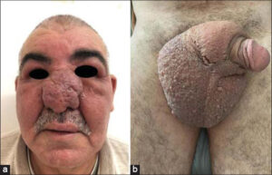 |
Figure 1: (a) Erythema, edema on both his eyelids, forehead, glabella and cheeks, also a rhinophyma nose. (b) Edema of the scrotum and scrotal lymphangiectasia. |
Consent
The examination of the patient was conducted according to the principles of the Declaration of Helsi
REFERENCES
1. Jędrowiak A, Oszukowska M, Żuchowska A, Tabara K, Szewczyk A, Kuchciak-Brancewicz M, et al. Penoscrotal elephantiasis:Case report. Our Dermatol Online. 2017;8:204-6.
2. Al Aboud A, Hakim M, Alehibi I, Alotaibi H, Al Saadi F, Bindogji Y, et al. Saxophone penis associated with penoscrotal lymphangiomatosis. Our Dermatol Online. 2022;13:e62.
Notes
Request permissions
If you wish to reuse any or all of this article please use the e-mail (brzezoo77@yahoo.com) to contact with publisher.
| Related Articles | Search Authors in |
|
|
|

Comments are closed.