Giant plexiform neurofibroma of the eyelid
Sara El-Ammari , Zakia Douhi, Imane Kacimi Alaoui, Hanane Baybay, Sara Elloudi, Meryem Soughi, Fatima-Zahra Mernissi
Department of Dermatology, University Hospital Hassan II, Fes, Morocco
Citation tools:


Copyright information
© Our Dermatology Online 2023. No commercial re-use. See rights and permissions. Published by Our Dermatology Online.
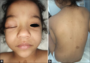
A 3-year-old girl with a history of delayed acquisition consulted for an asymptomatic mass of the right upper eyelid, observed by the mother since the age of 4 months, which had progressively increased in size. Physical examination revealed a 3 cm mass taking the right upper eyelid, of soft consistency with ptosis (Fig. 1a). The child also presented multiple axillary lentigines and more than 6 cafe au lait spots exceeding 5 mm in long axis on the trunk and limbs (Fig. 1b). A cerebro-orbital MRI confirmed that it was a plexiform neurofibroma of the right eyelid associated with dysplasia of the large right sphenoidal wing and glioma of the optic pathways, then she was referred to the ophthalmology department for management.
Neurofibromatosis type 1 (NF-1) is an autosomal dominant neurocutaneous disease. Plexiform neurofibromas (PNs) is pathognomonic of NF-1 and represents a rare kind of this disease where the neurofibroma originates from nerve sheath cells or subcutaneous peripheral nerves [1]. They are often congenital, but they may not be evident immediately after birth, they can increase rapidly in size before the age of 8 years, and they present a risk of malignant transformation [2]. PNs mostly occurs on the trunk and proximal extremities [3], they may be seen on the eyelids but are usually small size. The palpebral mass can be either firm or soft, sometimes the palpation of the involved tissues is said to resemble a ‘bag of worms’ and can result in ptosis [2]. At this location they tend to infiltrate the orbit, leading to severe proptosis and facial deformity [1], hence the interest in performing a cerebral and orbital MRI when a PN is suspected. Early surgical treatment before the age of 10 years may be necessary for a better functional and esthetic prognosis [2].
Consent
The examination of the patient was conducted according to the principles of the Declaration of Helsinki.
REFERENCES
1. Lam H, Harun Nor Rashid S. A case series of plexiform neurofibroma:the unusual presentations and surgical challenges. Cureus. 2022;14:e23141.
2. Avery RA, Katowitz JA, Fisher MJ, Heidary G, Dombi E, Packer RJ, et al. Orbital/Periorbital Plexiform Neurofibromas in Children with Neurofibromatosis Type 1:Multidisciplinary Recommendations for Care. Ophthalmology. 2017;124:123-32.
3. El Jouari O, Gallouj. S, Zinoune. S, Baybay. H, Mernissi FZ. Giant neurofibroma:a localization palpebral. Our dermatology online. 2018;9:329-30.
Notes
Request permissions
If you wish to reuse any or all of this article please use the e-mail (brzezoo77@yahoo.com) to contact with publisher.
| Related Articles | Search Authors in |
|
|
 http://orcid.org/0000-0001-6330-2856http://orcid.org/0000-0003-0582-321Xhttp://orcid.org/0000-0003-3455-3810http://orcid.org/0000-0002-5942-441X http://orcid.org/0000-0001-6330-2856http://orcid.org/0000-0003-0582-321Xhttp://orcid.org/0000-0003-3455-3810http://orcid.org/0000-0002-5942-441XMeryem Soughi
|

Comments are closed.