Palpebral bluish nodule: Think about hematoma
Sabrina Oujdi , Meryem Soughi, Siham Boularbah, Hanane Baybay, Sara Elloudi, Zakia Douhi, Fatima Zahra Mernissi
Department of Dermatology, University Hospital Hassan II, Fes, Morocco
Citation tools:


Copyright information
© Our Dermatology Online 2023. No commercial re-use. See rights and permissions. Published by Our Dermatology Online.
Subcutaneous hematoma is rare. It occurs spontaneously without bruising or following minimal bruising. They can be related to diseases responsible for coagulation disorders (such as hemophilia, leukemia, certain infectious diseases or liver failure). They can also occur when taking certain medications: anticoagulants, antiplatelet agents such as aspirin, corticosteroids taken over an extended period, cancer chemotherapy.
Hematomas under the skin appear as bluish lesions swollen with blood. They result from the rupture of larger blood vessels than those involved in the bruising.
Depending on their location, hematomas may be accompanied by other signs. When the eyelid is affected and the local swelling is significant, it can go as far as closing the eye completely and preventing vision [1]. The diagnosis of chronic subcutaneous hematoma can be mistaken for other pathologies such as basal cell carcinoma or pilomatricoma, we describe a new case of chronic subcutaneous hematoma.
A 74-year-old man with no notable pathological antecedents and no history of medication or previous trauma reports a sub-orbital nodule that was asymptomatic. On clinical examination presence of a firm subcutaneous nodule 0.5 cm long and painless at the level of the lower eyelid (Fig. 1a). Dermoscopy revealed the presence of a homogeneous blue pattern with fine telangiectasias (Fig. 1b). In the incision of the lesion presence of blood clot without underlying lesions.
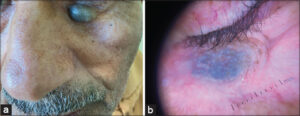 |
Figure 1: (a) Clinical picture: blue nodule under palpebral. (b) Dermoscopic picture: homogeneous blue pattern with fine telangiectasias. |
Consent
The examination of the patient was conducted according to the principles of the Declaration of Helsinki.
REFERENCE
1. Estève E, Armingaud P, Martin L. Hématomes cutanés et sous-cutanés vus en dermatologie:17 cas. Ann Dermatol Venereol. 2004;131:555-8.
Notes
Request permissions
If you wish to reuse any or all of this article please use the e-mail (brzezoo77@yahoo.com) to contact with publisher.
| Related Articles | Search Authors in |
|
|
 http://orcid.org/0009-0002-5084-9152http://orcid.org/0000-0002-3975-8316http://orcid.org/0000-0003-3455-3810http://orcid.org/0000-0002-5942-441X http://orcid.org/0009-0002-5084-9152http://orcid.org/0000-0002-3975-8316http://orcid.org/0000-0003-3455-3810http://orcid.org/0000-0002-5942-441X |

Comments are closed.