Atypical nodular BCC of the eyelid
Imane Couissi , Sara El Loudi, Zineb Fajri, Meryem Soughi, Zakia Douhi, Hanane Baybay, Fatima Zahra Mernissi
Department of Dermatology, University Hospital Hassan II Fès, Morocco
Citation tools:


Copyright information
© Our Dermatology Online 2023. No commercial re-use. See rights and permissions. Published by Our Dermatology Online.
Among skin cancers, basal cell carcinomas (BCCs), are the most common type, representing 80% of all non-melanoma malignant skin tumors [1]. More than 80% occur in the head and neck region [2], followed by the trunk (15%) and extremities (only 5%).
Of all BCCs detected in the head and neck region, 14% grow in the periocular area, half of them being encountered on the lower eyelid and a quarter in the medial canthus. The lateral canthus and upper lid are the least involved.
In the eyelid location, the three most common histological subtypes are the morphea form, infiltrative, and basosquamous subtypes, which constitute over 80% of BCC cases with orbital invasion [3].
We describe a case of nodular BCC of the upper eyelid histologically confirmed.
A 42-year-old man presented with a 2-year history of a nodular lesion on the tail of the right that has progressively increased in size.
Clinical examination revealed a well-limited 2cm erythematous nodule, with a hard consistency fixed in relation to the deep plane in some places, resting on an atrophic erythematous plaque with a pearly border in some places, located at the level of the right eyebrow tail (Fig. 1a). Dermoscopy shows erythematous background, telangiectasias, and tree trunk vessels (Figs. 1b and 1c).
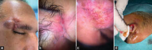 |
Figure 1: (a) A well-limited 2cm erythematous nodule, with a hard consistency fixed in relation to the deep plane in some places, resting on an atrophic erythematous plaque with a pearly border in some places, located at the level of the right eyebrow tail. (b and c) Dermoscopy showing erythematous background, telangiectasias, and tree trunk. (d) Intraoperative exploration during the biopsy showing a black pigment. |
The examination of the lymph nodes was free.
Intraoperative exploration during the biopsy showed a black pigment (Fig. 1d). The result of the biopsy was in favor of a pigmented nodular BCC (Fig. 2). Complete surgical excision was performed with margin and covering.
 |
Figure 2: Histological image showing a basaloid appearance in the tumor cells. The nuclei have a palisading arrangement with the presence of retraction slits (HESx100/200). |
Consent
The examination of the patient was conducted according to the principles of the Declaration of Helsinki.
REFERENCES
1. Sarma DP, Olson D, Olivella J, Harbert T, Wang B, Ortman S. Clear cell basal cell carcinoma. Patholog Res Int. 2011.2011:386921.
2. Shi Y, Jia R, Fan X. Ocular basal cell carcinoma:a brief literature review of clinical diagnosis and treatment. Onco Targets Ther. 2017;10:2483–9.
3. Sun MT, Wu A, Figueira E, Huilgol S, Selva D. Management of periorbital basal cell carcinoma with orbital invasion. Future Oncol.2015;11:3003–10.
Notes
Request permissions
If you wish to reuse any or all of this article please use the e-mail (brzezoo77@yahoo.com) to contact with publisher.
| Related Articles | Search Authors in |
|
|
 http://orcid.org/0009-0005-2261-6919http://orcid.org/0000-0002-5942-441Xhttp://orcid.org/0000-0003-3455-3810 http://orcid.org/0009-0005-2261-6919http://orcid.org/0000-0002-5942-441Xhttp://orcid.org/0000-0003-3455-3810 |

Comments are closed.