Basal cell carcinoma of the scalp mimicking superficial granulomatous pyoderma
Cyrine Marmech 1, Fouzia Hali1, Farida Marnissi2, Soumiya Chiheb1
1Department of Dermatology, CHU Casablanca, Morocco, 2Department of Pathological Anatomy, CHU Casablanca, Morocco
Citation tools:


Copyright information
© Our Dermatology Online 2023. No commercial re-use. See rights and permissions. Published by Our Dermatology Online.
Basal cell carcinoma (BCC) is one of the most common neoplastic proliferations. BCC is locally aggressive [1]. Its evolution may mimic superficial granulomatous pyoderma (SGP). The diagnosis is clinical and histological. Herein, we report a case of BCC of the scalp diagnosed by dermoscopy and confirmed by histology.
A 69-year-old patient consulted for an ulcerated, painful scalp evolving for ten years (Fig. 1) A medical examination found an ulcer-budding patch, 20 × 10 cm in size, some healing areas, a raised border, an irregular edge, some residual occipital hair. A dermoscopic examination revealed large, tree-like vessels, fine ramifications, telangiectasias, and a well-defined, ovoid structure with bluish-gray pigmentation (Figs. 2a and 2b). A histological study concluded to infiltrating BCC. A craniofacial CT scan found an erosion in the right frontal bone. A thoraco-abdomino-pelvic CT scan revealed no secondary localizations. Radiotherapy was indicated as a treatment.
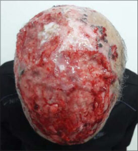 |
Figure 1: Ulcer-budding patch on the scalp. |
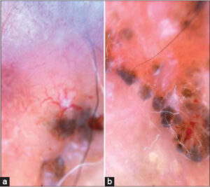 |
Figure 2: (a-b) Large, tree-like vessels, fine ramifications, telangiectasias, and a well-defined, ovoid structure with bluish-gray pigmentation. |
The giant ulcer-budding aspect of the whole scalp makes superficial granulomatous pyoderma (PGS) clinically discussable. Dermoscopic examination, especially the fine telangiectasias, the large tree-like vessels with fine ramifications, the bluish-gray, pigmented, ovoid structures, was in support of CBC, which was confirmed histologically. Ovoid nests are commonly found in both CBC and PGS. The existence of pustules (missing in our case) would be suggestive of pyoderma [2], whereas the other dermoscopic elements are more likely to be indicative of CBC [3]. Surgical treatment would be difficult in this case.
Dermoscopy before biopsy helps to guide the diagnosis of challenging cases confusing inflammatory and tumor pathology.
Consent
The examination of the patient was conducted according to the principles of the Declaration of Helsinki.
The authors certify that they have obtained all appropriate patient consent forms, in which the patients gave their consent for images and other clinical information to be included in the journal. The patients understand that their names and initials will not be published and due effort will be made to conceal their identity, but that anonymity cannot be guaranteed.
REFERENCES
1. Furukawa H, Sowa-Osako J, Ozawa T, Hashimoto T, Tsuruta D. A case of a long-neglected basal cell carcinoma on the scalp. Our Dermatol Online. 2021;12:206-7.
2. Rosina P, Papagrigoraki A, Colato C. A case of superficial granulomatous pyoderma mimicking a basal cell carcinoma. Acta Dermatovenerol Croat. 2014;22:48-51.
3. Laamari K, Douhi Z, Elloudi S, Baybay H, Mernissi FZ. Basal cell carcinoma: An unusual localization. Our Dermatol Online. 2020;11:e74.1-e74.2.
Notes
Request permissions
If you wish to reuse any or all of this article please use the e-mail (brzezoo77@yahoo.com) to contact with publisher.
| Related Articles | Search Authors in |
|
|
|

Comments are closed.