When dermatoscopy makes the diagnosis of facial atypical pigmentation
Massara Baklouti , Khadija Sellami, Mariem Rekik, Hamida Turki
Dermatology department, Hedi Chaker Hospital, Sfax University, Tunisia
Citation tools:


Copyright information
© Our Dermatology Online 2023. No commercial re-use. See rights and permissions. Published by Our Dermatology Online.
OBSERVATION
A 35-year-old female patient, with a history of hypothyroidism, presented with pruritic macular hyperpigmentation of the face and neck evolving for months. There were no other skin lesions. Histology revealed an orthokeratotic epidermis with a perifollicular lymphocytic infiltrate. There was also atrophy of the follicular sheaths with keratinocyte necrosis. The most likely diagnosis was pigmentosus lichen and she was put on dermocorticoids. There was no improvement after 2 months. Examination revealed poorly limited inhomogeneous pigmented macules of the forehead, temporal regions and neck with multiple open comedones (Fig. 1). The pigmentation enhanced at the follicular level becoming blackish. Dermatoscopy revealed horny black plugs with intense perifollicular pigmentation giving the appearance of black circles and black dots in a linear pattern without detectable vessels (Fig. 2a). The patient denied comedogenic exogenous topical use or specific occupational exposure. Cleansing with water removed a blackish pigment (Fig. 2b) and made this follicular pigmentation disappear on dermatoscopy, leaving a background of erythema with diffuse grayish pigmentation (Fig. 2c).
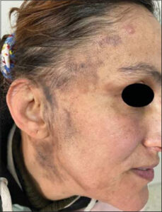 |
Figure 1: Pigmented macules with multiple comedones. |
 |
Figure 2: (a) On dermatoscopy: horny black plugs giving the appearance of black circles and dots in a linear pattern. (b) A blackish pigment is removed after cleansing. (c): Follicular pigmentation disappears. |
WHAT IS YOUR DIAGNOSIS?
Answer: Exogenous facial pigmentation. Chronic rubbing would be the cause of the residual hyperpigmentation. The patient was referred to a psychiatrist.
DISCUSSION
Facial hyperpigmentation is one of the most common chief complaints in aesthetic consultation [1]. Dermatoscopy is a diagnostic tool that is increasingly used in inflammatory dermatoses, particularly pigmented dermatoses [2]. The diagnosis of facial hyperpigmentation is often challenging because of the multitude of etiologies and clinico-pathological similarities. Herein, we report a misleading case of an unusual facial hyperpigmentation. Our case illustrates the important role of dermatoscopy in rectifying the diagnosis. It may reduce the need of invasive procedures [3]. Dermatitis artefacta is a challenging psychocutaneous disorder because of the clinical polymorphism. It should be evoked in case of chronic and recurrent dermatosis which can be induced or simply worsened [4]. Histologic changes in skin induced by friction are predominantly found around hair follicles and eccrine ducts. Factitious disorder should be considered if there is cell necrosis [5].
REFERENCES
1. Samson N, Fink B, Matts PJ. Visible skin condition and perception of human facial appearance. Int J Cosmet Sci. 2010;32:167-84.
2. Micali G, VerzìAE, Lacarrubba F. Alternative uses of dermoscopy in daily clinical practice:An update. J Am Acad Dermatol. 2018;79:1117-1132.e1.
3. Lacarrubba F, VerzìAE, Dinotta F, Scavo S, Micali G. Dermatoscopy in inflammatory and infectious skin disorders. G Ital Dermatol Venereol. 2015;150:521-31.
4. Chandran V, Kurien G. Dermatitis Artefacta. 2022 Jul 12. In:StatPearls. Treasure Island (FL):StatPearls Publishing;2022 Jan–.
5. Tittelbach J, Peckruhn M, Elsner P. Histopathological patterns in dermatitis artefacta. J Dtsch Dermatol Ges. 2018;16:559-64.
Notes
Request permissions
If you wish to reuse any or all of this article please use the e-mail (brzezoo77@yahoo.com) to contact with publisher.
| Related Articles | Search Authors in |
|
|
 http://orcid.org/0000-0002-7964-062Xhttp://orcid.org/0000-0002-0842-2414 http://orcid.org/0000-0002-7964-062Xhttp://orcid.org/0000-0002-0842-2414 |

Comments are closed.