Erythematous nodule of the arm
Najoua Ammar 1, Mariam Meziane1, Nadia Ismaili1, Leila Benzekri1, Kawtar Znati2, Karima Senouci1
1Department of Dermatology and Venereology, CHU Ibn Sina, Université Mohammed V, Rabat, Morocco, 2Department of Pathological Anatomy, Ibn Sina Hospital, Rabat, Morocco
Citation tools:


Copyright information
© Our Dermatology Online 2023. No commercial re-use. See rights and permissions. Published by Our Dermatology Online.
Sir,
Eccrine poroma is a rare benign tumor of adults that shares the broad topography of eccrine sweat glands.
We report the observation of a case of eccrine poroma unusual by its location in the arm
A 63-year-old patient, with no previous pathological history, consulted for a tumor located on the left arm, evolving for 5 years, which had recently become painful and hemorrhagic.
The dermatological examination revealed a nodular erythematous tumor with a peripheral pigmented border on the inner side of the right arm, with a firm consistency. The tumor measured 3.5 cm in long axis and was sessile (Fig. 1a).
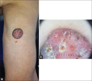 |
Figure 1: (a) Nodular erythematous tumor with a peripheral pigmented border on the inner side of the right arm. (b) Dermoscopy showing polymorphic vascularization: areas without pink structure, tree-like vessels, in globi and in leaves. |
The dermosocopy showed pink areas without any structure containing vessels called leaves surrounded by whitish avascular structures (Fig. 1b).
Biopsy of the tumor was performed. The skin histology showed an epithelial proliferation emanating from the basal layer of the epidermis made of regular cells, with basophilic nuclei, arranged in compact trabeculae.
All these elements were in favor of an eccrine poroma.
A total excision of the lesion was performed.
Eccrine poroma is a benign adnexal tumor described in 1956 by Pinkus. It represents 10% of the tumors of the sweat glands. It is observed in adults between 40 and 60 years of age, without gender predominance.
Its pathophysiology is poorly understood; however, factors such as repeated exposure to radiation [1] and trauma have been incriminated. Typically, it presents as a single papule, plaque, or nodule that slowly evolves into a fleshy tumor that may ulcerate [1]. These tumors are budding, fleshy, and preferentially localized to the pre-calcifixion plantar regions. Dermoscopy can be helpful in eliminating differential diagnoses. The appearance of “leafy” or “flowering” vessels is suggestive [1,2]. It is shown by epithelial trabeculae developed from the basal layer, well limited in relation to the surrounding epidermis. Ductal differentiation is demonstrated by periodic Schiff’s acid (PAS) and immunohistochemistry with antibodies against carcinoembryonic antigen (CEA) or epithelial membrane antigen (EMA) [3].
Treatment is surgical and excisionl must be complete. The clinical course of our patient was favorable and no recurrence was noted after a 5-month follow-up.
Our observation is peculiar for the unusual location of an eccrine poroma in the arm.
Consent
The examination of the patient was conducted according to the principles of the Declaration of Helsinki.
The authors certify that they have obtained all appropriate patient consent forms, in which the patients gave their consent for images and other clinical information to be included in the journal. The patients understand that their names and initials will not be published and due effort will be made to conceal their identity, but that anonymity cannot be guaranteed.
REFERENCES
1. McCoskey M, Neerukonda VK, Hatton MP, Wolkow N. Eccrine poroma of the eyelid. Orbit. 2021 Jul 14:1.
2. Chessa MA, Patrizi A, Baraldi C, Fanti PA, Barisani A, Vaccari S. Dermoscopic-Histopathological Correlation of Eccrine Poroma:An Observational Study. Dermatol Pract Concept. 2019;9:283-91.
3. Mohamed AM, Mohamed BJ, Rania K, Montassar G. Eccrine poroma localized in the second toe. Radiol Case Rep. 2022;17:4108-10.
Notes
Request permissions
If you wish to reuse any or all of this article please use the e-mail (brzezoo77@yahoo.com) to contact with publisher.
| Related Articles | Search Authors in |
|
|
 http://orcid.org/0000-0001-6348-4450 http://orcid.org/0000-0001-6348-4450 |

Comments are closed.