Atypical distribution of lichen planus pigmentosus: Pigmentation of the nose and ears
Chaymae Jroundi , Hanane Baybay, Siham Boularbah, Zakia Douhi, Sara Elloudi, Fatima Zahra Mernissi
Department of Dermatology, University Hospital Hassan II, Fes, Morocco
Citation tools:


Copyright information
© Our Dermatology Online 2023. No commercial re-use. See rights and permissions. Published by Our Dermatology Online.
Sir,
Lichen planus pigmentosus (LPP) is considered a rare variant of lichen planus (LP). It typically presents with a symmetrical distribution of dark brown to grayish-blue macules and blotches, which eventually enlarge and coalesce. Usually localized on the face, it has a predilection for the temporal areas, whereas on the neck, it affects all sides [1]. There are some reported cases of a linear pattern, a zosteriform pattern over the trunk, the involvement of non-sun exposed areas, such as the thighs, and a band-like distribution of LPP on the abdomen [2]. Dermoscopy in LPP demonstrates pigmented globules, whose color ranges from dark brown to bluish-gray, arranged in diffuse, dotted, annular, hem-like, arcuate, speckled, and perifollicular patterns. Wickham striae and vascular features are absent, unlike in classical lichen planus [3,4]. Histopathology reveals a vacuolar interface dermatitis with band-like lichenoid or perivascular lymphocytic infiltrates in the papillary dermis, as well as superficial pigmentary incontinence and melanophages [3,5]. The differential diagnoses are melasma, ashy dermatoses, often present on the trunk and limbs, ochronosis, and post-inflammatory hyperpigmentation. The treatment of LPP has had limited success and there is no clear consensus. Topical treatment includes corticosteroids, tacrolimus, hydroquinone, and retinoids. One of the most commonly employed topical treatments is tacrolimus. Refractory forms may be treated with systemic corticosteroids with gradual tapering, as well as dapsone or isotretinoin [5]. Herein, we report a case of an atypical clinical distribution of LPP with a typical dermoscopic pattern.
A 71-year-old male patient visited our department for a hyperpigmentation of the nose and the auricles of the ears evolving for a month. He had no history of medication or the application of hydroquinone or other topical ointments. A clinical examination revealed well-limited hyperpigmented macules with irregular borders located on the tip of the nose and the auricle of both ears (Figs. 1a and 1b). This atypical clinical distribution of lesions along cartilaginous zones was initially suggestive of alkaptonuria. The diagnosis was less likely, given the absence of black discoloration of the urines and the absence of pigmentation of the sclera. Dermoscopy revealed brown and gray globules arranged in a granular, annular, perifollicular pattern, which was more suggestive of LPP despite the atypical topography (Fig. 2). The diagnosis of LPP was confirmed by histopathology, which showed an interface dermatitis with a lichenoid infiltrate of the dermis consisting of lymphocytes and plasma cells with numerous melanophages. The atypical distribution on the ears might have be related to the Koebner’s phenomenon as the patient tended to wear glasses. Yet, this does not explain the location on the tip of the nose. The patient was treated with topical steroids with good evolution.
 |
Figure 1: (a) Clinical presentation: well-limited, hyperpigmented macules with irregular borders located on the tip of the nose. (b) Clinical presentation: well-limited, hyperpigmented macules with irregular borders located on the tip of the auricle of the ear. |
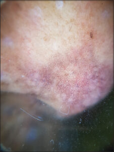 |
Figure 2: Dermoscopy of the nose: brown and gray globules arranged in a granular, annular, perifollicular pattern. |
Consent
The examination of the patient was conducted according to the principles of the Declaration of Helsinki.
The authors certify that they have obtained all appropriate patient consent forms, in which the patients gave their consent for images and other clinical information to be included in the journal. The patients understand that their names and initials will not be published and due effort will be made to conceal their identity, but that anonymity cannot be guaranteed.
REFERENCES
1. Ghosh A, Coondoo A. Lichen planus pigmentosus:The controversial consensus. Indian J Dermatol. 2016;61:482-6.
2. Nag F, Ghosh A, Chatterjee G, Choudhary N. Lichen planus pigmentosus:Two atypical presentation. Our Dermatol Online. 2013;4:78-9.
3. Ankad BS, Drago NR, Koti VR, Nikam BP. Dermoscopic approach to hyperpigmented lesions in skin of color. Clin Dermatol Rev. 2020;4:84-91.
4. El Jouari O, Senhaji G, Gallouj S, Baybay H, Mernissi FZ. Lichen planus pigmentosus. Our Dermatol Online. 2019;10:404-5.
5. Robles-Méndez JC, Rizo-Frías P, Herz-Ruelas ME, Pandya AG, Ocampo Candiani J. Lichen planus pigmentosus and its variants:Review and update. Int J Dermatol. 2018;57:505-14.
Notes
Request permissions
If you wish to reuse any or all of this article please use the e-mail (brzezoo77@yahoo.com) to contact with publisher.
| Related Articles | Search Authors in |
|
|
 http://orcid.org/0000-0003-3455-3810http://orcid.org/0000-0002-3975-8316http://orcid.org/0000-0002-5942-441X http://orcid.org/0000-0003-3455-3810http://orcid.org/0000-0002-3975-8316http://orcid.org/0000-0002-5942-441X |

Comments are closed.