Koplik’s spots
Abdullah Mancy
Private Clinic of Dermatology and Venereology, Al-Ramadi City, Al-Anbar Governorate, Iraq
Citation tools:


Copyright information
© Our Dermatology Online 2023. No commercial re-use. See rights and permissions. Published by Our Dermatology Online.
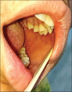
Koplik’s spots are pathognomonic features of measles in its prodromal phase. They are named after American pediatrician Dr. Henry Koplik, who first described them in 1896. They appear 2–3 days before the onset of a measles rash as small, bluish-white, slightly elevated papules on erythematous bases on the buccal mucosa, usually opposite the first and second lower molar teeth. Because of their characteristic appearance, they are described as “grains of salt on a reddish background.” They may spread to involve other parts of the buccal cavity, pharynx, and soft palate. Occasionally, they occur on the conjunctiva and the vaginal and gastrointestinal mucosae. The white color of the spots may result from the destruction of cells of the glandular epithelium. They persist for 12 hours to 4 days and fade on the rash appearance. When the skin rash appears and progresses, the spots lose their characteristic appearance, and after several days, the mucosa returns to its normal appearance [1,2].
A fifteen-year-old child presented with high fever, tiredness, conjunctival congestion, and upper respiratory tract symptoms persisting for the past two days. On examination of the mouth, there were multiple, white spots on erythematous bases facing the upper and lower molar teeth (Fig. 1). The patient was diagnosed with a case of measles and admitted to the hospital for further investigations and treatment.
Consent
The examination of the patient was conducted according to the principles of the Declaration of Helsinki.
The authors certify that they have obtained all appropriate patient consent forms, in which the patients gave their consent for images and other clinical information to be included in the journal. The patients understand that their names and initials will not be published and due effort will be made to conceal their identity, but that anonymity cannot be guaranteed.
REFERENCES
1. Brzezinski P, Fiorentino DF, Arunachalam P, Katafigiotis I, Matuszewski Ł, Narita M, et al. Dermatology eponyms –sign –lexicon –(K). Our Dermatol Online. 2014;5:95-102.
2. Jain SA, Rao MS, Mahesh AR. Blue in dermatology. Our Dermatol Online. 2022;13:e6.
Notes
Request permissions
If you wish to reuse any or all of this article please use the e-mail (brzezoo77@yahoo.com) to contact with publisher.
| Related Articles | Search Authors in |
|
|
 http://orcid.org/0000-0002-3688-6953 http://orcid.org/0000-0002-3688-6953 |

Comments are closed.