Diagnostic wandering of a case of giant pedicled lipoma of the left inguinal fold
Mamadou Maïmouna Ouédraogo1, Laouali Salissou 1, Laouali Idi Mamane Sani1, Alban Michel Bassolé2, Mariama Abdoulaye1, Muriel Ouédraogo2, Yaya Ouedraogo2, Amina Nongtondo2, Patrice Tapsoba2, Nessiné Nina Korsaga/Somé2, Fatou Barro2, Pascal Niamba2, Adama Traoré2
1Department of Dermatology Venereology, National Hospital of Niamey, Niger, 2Department of Dermatology Venereology, CHU YO Ouagadougou, Burkina Faso
Citation tools:


Copyright information
© Our Dermatology Online 2023. No commercial re-use. See rights and permissions. Published by Our Dermatology Online.
ABSTRACT
Lipomas are usually benign tumors formed from a proliferation of mature adipocytes, resulting in hypodermic, soft, compressible, and mobile nodular formations under the skin. In their subcutaneous location, superficial lipomas represent 16% to 50% of soft tissue tumors. They may be solitary or multiple. Solitary lipoma is usually seen in young adults between the ages of 30 and 50 years, regardless of sex, and is frequently asymptomatic. A lipoma is called giant when its weight exceeds 1 kg or its diameter exceeds 5 cm. The etiopathogenesis of lipomas is poorly understood. Herein, we report a case of giant pedunculated lipoma localized on the left inguinal fold being a distress for the patient.
Key words: Giant pedunculated lipoma, Diagnostic wandering, Ablation, Burkina Faso
INTRODUCTION
Lipomas are generally benign tumors formed from a proliferation of mature adipocytes, resulting in hypodermic, soft, compressible, and mobile nodular formations under the skin. They are most often encapsulated and slow-growing, reaching large dimensions. Usually sessile, in rare conditions, a lipoma is referred to as giant when its weight exceeds 1 kg or its diameter exceeds 5 cm [1–3]. Herein, we report a case of giant lipoma localized on the left inguinal fold being a distress for the patient.
CASE REPORT
This was a forty-year-old patient, farmer, residing in Djibo, Burkina Faso, monogamous, father of four children, with no known pathological history. He consulted for an asymptomatic mass localized on the left inguinal fold evolving for ten previous years and distressing the patient.
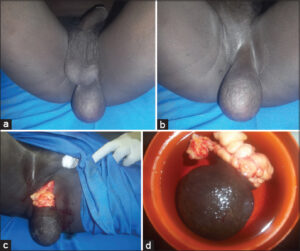
A physical examination revealed a tumor lesion, 5–12 cm in size, soft, non-depressible, mobile with respect to the deep plane, and superficial, localized on the left inguinal fold (Fig. 1a), pedunculated on a base of 4 cm, not painful on palpation. The skin in the front was normal. Another tumor lesion molasse (molluscum pendulum) of 1–2 cm was on the inner surface of the right buttock (Fig. 1b). An ultrasound examination confirmed that it was an avascular mass characteristic of a lipoma. For management, we performed a surgical excision (Fig. 1c), during which we, on gross examination, discovered encapsulated fatty lobules, confirming the diagnosis of giant pedunculated lipoma, whose weight was 830 g (Fig. 1d). Electrocoagulation of the small tumor (molluscum pendulum) was performed. An anatomopathological examination of the room confirmed that it was a lipoma.
DISCUSSION
In their subcutaneous localization, superficial lipomas account for 16% to 50% of soft tissue tumors. They may be solitary or multiple. Solitary lipoma is generally seen in young adults between 30 and 50 years of age, regardless of sex, and is frequently asymptomatic [3,4], as in our patient, yet sometimes, it is painful [5]. The complaint in the latter was essentially functional discomfort and anxiety about the impact on his libido and fertility. The diagnosis is guided by clinical examination, as in our patient. Lipoma is often painless and usually results in a soft, regular, mobile tumor. It is located on the back in 15% to 20% of cases yet may be localized anywhere on the body [1,2,6]. The clinical presentation of the lesion in our patient (shape, size, pedunculated appearance, location) was not usual in lipomas and may be explained by the permanent pressure exerted by the inguinal fold and the action of gravity on the lesion. Posch described the clinical test of ice application to the tumor, which in the case of lipoma, results in the solidification of the mass. The usual course is slow growth, which may stabilize spontaneously [2,3]. The soft consistency and pedunculated appearance at first evoked molluscum pendulum, similarly to our daily practice, or inguinal hernia [2], yet the non-depressible aspect such as a hollowed out grape grain was not in favor. Differential diagnosis was also made with other soft tissue tumors such as angio-eccrine hamartoma [7] and hidradenoma [8].
Some authors opt for primary liposuction to reduce tumor volume, because the aesthetic result would be more satisfactory and post-operative morbidity would be decreased [3,6].
CONCLUSION
Our patient made a diagnostic wandering of ten years despite his numerous consultations. Abstention from treatment was the choice in previous consultations because of the location and the unusual clinical appearance of the lesion. This was a source of major anxiety and moral prejudice in our patient. Surgical treatment is of choice with total remission.
Consent
The examination of the patient was conducted according to the principles of the Declaration of Helsinki.
The authors certify that they have obtained all appropriate patient consent forms, in which the patients gave their consent for images and other clinical information to be included in the journal. The patients understand that their names and initials will not be published and due effort will be made to conceal their identity, but that anonymity cannot be guaranteed.
REFERENCES
1. Niasse A, Faye PM, Ndong A, Thiam O, Gueye O, Gueye ML, et al. [Giant lipoma of the back:A case report and literature review]. Pan Afr Med J. 2022;42:292.
2. Yuksel ME, Tamer F, Oz E. A giant groin lipoma mimicking an inguinal hernia:A case report. Our Dermatol Online. 2019;10:38-40.
3. Sboui I, Riahi H, Jlalia Z, Daghfous MS, Chelly-Bouaziz M. Parosteal lipoma of the lower limb:Report of two cases. Ann Orthop Musculoskelet Disord. 2018;1:10-3.
4. Abdullah M, Lateef A. Nevus lipomatosus superficialis. Our Dermatol Online. 2020;11:412.
5. Fnini S, Hassoune J, Largab A. Lipome géant de la main. Rev Chir Main. 2010;29:44-7.
6. Murphey MD, Carroll JF, Fleming DJ. Lipomatous lesions. Radiographics. 2004;24:1433-66.
7. KantéMD, Diop K, Ngom NB, Andriateloasy S, Deh A, Ndiaye M, et al. Angio-eccrine hamartoma:About two cases in Dakar, Senegal. Our Dermatol Online. 2022;13(Supp. 2):45-8.
8. Marraha F, Al Faker I, Chahoub H, Benyamna Y, Rahmani N, Salim G. Hidradenoma with an atypical localization mimicking lipoma. Our Dermatol Online. 2022;13:469-70.
Notes
Request permissions
If you wish to reuse any or all of this article please use the e-mail (brzezoo77@yahoo.com) to contact with publisher.
| Related Articles | Search Authors in |
|
|
 http://orcid.org/0000-0002-0734-0759 http://orcid.org/0000-0002-0734-0759 |

Comments are closed.