Rickettsial diseases: A group of underdiagnosed fevers
Kacimi Alaoui Imane , Zakia Douhi, Sara El-Ammari, Meryem Soughi, Sara Elloudi, Hanane Baybay, Fatima-Zahra Mernissi
Department of Dermatology, University Hospital Hassan II, Fes, Morocco
Citation tools:


Copyright information
© Our Dermatology Online 2023. No commercial re-use. See rights and permissions. Published by Our Dermatology Online.
ABSTRACT
Background: Rickettsias are zoonoses transmitted to humans by arthropods. Their clinical manifestations vary. Prompt and effective treatment is recommended before biological confirmation. The aim of this study was to establish the epidemiological and clinical profiles of this group as well as its different extracutaneous manifestations.
Materials and Methods: This retrospective study included patients with rickettsiosis who consulted between June 2016 and October 2022.
Results: Twenty-nine patients, with a male predominance of 66%, aged between 2 and 74 years. A maculopapular rash was present in all cases. Symptomologies ranged from neurological and digestive to renal. All our patients underwent a rickettsia check-up and serology. Twenty-five patients presented a febrile cutaneous eruption forty-eight hours after the tick, leading to hospitalization and prompt treatment. Evolution was characterized by disinfiltration and positive improvement.
Conclusion: Because rickettsial diseases have a wide geographical distribution, recognition of their symptoms is essential for prompt treatment.
Key words: Rickettsias; Eruption; Escharotic
INTRODUCTION
Tick-borne rickettsia is caused by Gram-negative bacteria belonging to the spotted fever group of the genus Rickettsia [1]. Among the oldest vector-borne diseases, these zoonotic diseases are the most common. The first clinical description of Rocky Mountain spotted fever (RMSF) in Idaho, U.S., was reported by Edward E. Maxey in 1899, and the first case of Mediterranean spotted fever (MSF) was reported by Conor and Brush in Tunisia in 1910 [2]. The disease affects people of all ages and mainly occurs in summer. The clinical presentation varies from mild to highly severe, with a mortality of 2–6% [3]. The signs are fever, headache, maculopapular rash, an escharotic spot, and localized adenopathy. Rickettsial diseases are also difficult to diagnose, as there are no rapid point-of-care tests to establish the diagnosis during acute infection, and the confirmation of the diagnosis, when sought, is usually retrospective using serological methods [3]. Treatment should be administered prior to biological confirmation for early and rapid improvement [4]. The objective of our study was to identify the epidemiological and clinical characteristics of our patients followed for rickettsial disease.
MATERIALS AND METHODS
In this prospective, retrospective, cross-sectional study, we collected all cases of rickettsiosis consulted at Hassan II Hospital of Dermatology and the emergency room between June 2016 and October 2022.
RESULTS
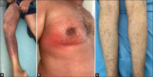
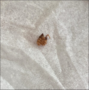
We collated 29 patients, with a male predominance of 66%, aged between 2 and 74 years. The legs and thighs were the most frequent topography of the escharotic spot (Fig. 1a); maculopapular rash was present in all our cases (Figs. 1b and 1c); purpura was present in 67%. The extracutaneous symptoms were rich and varied between neurological in 7 patients (headache, dizziness, behavioral, and consciousness disorders), digestive in 5 (abdominal pain, nausea, and vomiting), and renal in 10. The average time to onset of the symptoms was 4 days. Six patients were admitted to the emergency room for a disorder of consciousness diagnosed late as secondary to rickettsiosis. Biological tests showed hyperleukocytosis with a neutrophil predominance and elevated CRP in all our patients. Serology for rickettsial disease was positive in twelve patients. Our patients described a typical cutaneous reaction composed of a febrile cutaneous and mucosal rash forty-eight hours after the tick bite (Fig. 2), particularly in one patient, who presented a declining infiltrated purpura on the limbs, diagnosed as vasculitis after a cutaneous biopsy. Doxycycline 200 mg/day was prescribed as a formal indication. A lumbar puncture and cerebral MRI of four patients revealed secondary cerebral MRI of rickettsial disease. There was also vasculitis and encephalitis. In the case of abdominal pain and lipasemia five times normal, an abdominal scan was performed, which showed pancreatitis at stage E, from where the indication of the addition of ciprofloxacin for seven days was given to the six patients. The evolution was marked by the disinfiltration of the rash and the healing of the escharotic ulceration, as well as the improvement of other cerebral and digestive conditions. However, one patient ultimately died. This was due to his late consultation and the late initiation of therapy.
DISCUSSION
Rickettsia, also known as spotted fever, is caused by Rickettsia conorii and is transmitted by the brown dog tick [5]. Cases occur during the summer months when ticks are most active [6].
The time from the moment of infection and the incubation period to the onset of the disease is six days on average. Often, patients present with a sudden increase in body temperature, flu-like syndrome (headache, chills, joint, and muscle pain), and scabs (“black spots”) at the site of the tick bite [7]. These scabs are characteristic of the disease. These are red, inflamed papules, necrotic in the center, black, painless, more often located on the trunk, legs, or arms (in infants, they are more often found on the scalp in the occipital region). Sometimes, scabs may be absent. A generalized maculopapular rash (97%) affects the extremities and trunk. Other common clinical symptoms include myalgia, headache, conjunctivitis, hepatomegaly, and splenomegaly. Gastrointestinal symptoms may occur in approx. 30% of patients and are more common in children [8]. The severe form occurs in 5–6% of cases and is associated with disseminated vasculitis, renal, neurological, and cardiovascular complications, and phlebitis. In a recent prospective study in Algeria, 49% of patients were hospitalized with severe forms. The overall mortality rates for patients hospitalized with severe neurologic signs and multiple lesions were 3.6% and 54.5% [9]. In order to include it in the differential diagnosis of undifferentiated fever, the clinical symptoms and epidemiological knowledge of rickettsia are required. Serology is the basis for diagnosis and indirect immunofluorescence (ELISA) is the serological method of choice [10]. If rickettsiosis is suspected, empirical treatment should be initiated prior to in vitro laboratory susceptibility and in vivo confirmation. Doxycycline is currently recommended for the treatment of rickettsiosis. For adults, doxycycline 200 mg for 2-to-5 days or within twenty-four hours of fever is most commonly used, yet doxycycline 200 mg once administered has been shown to be effective in some cases of rickets in the spotted fever group [11]. The treatment of severe forms of the disease involves an intravenous administration of 200 mg doxycycline per day, followed by oral administration of 200 mg doxycycline per day until complete recovery (10 days). For children and pregnant women, treatment with certain macrolides for 5 to 7 days is recommended. However, a single dose of doxycycline at 5 mg/kg/day is effective and does not cause the side effect of tooth discoloration. In individuals allergic to tetracycline, ciprofloxacin (1.5 g/day, orally administered for 5 days) or chloramphenicol (2 g/day, 7 to 10 days) is effective against rickettsia erythematosus [12]. Its prevention relies on early detection (within twenty hours) and the appropriate removal of ticks in order to avoid transmission. There are no vaccines available for the prevention of rickettsial diseases or antibiotic prophylaxis. We have outlined several points concerning this bacterial infection in our cases and the literature: it is a severe infection that is potentially fatal, and dermatologists should be aware of it so that they may initiate TRT without delay.
CONCLUSION
Rickettsial diseases are present worldwide and continue to emerge and re-emerge as leading causes of febrile illness. Early identification of clinical presentations will be helpful in prompt and appropriate treatment.
Statement of Human and Animal Rights
All the procedures followed were in accordance with the ethical standards of the responsible committee on human experimentation (institutional and national) and with the 2008 revision of the Declaration of Helsinki of 1975.
Statement of Informed Consent
Informed consent for participation in this study was obtained from all patients.
REFERENCES
1. Parola P, Paddock CD, Raoult D. Tick-borne rickettsioses around the world:Emerging diseases challenging old concepts. Clin Microbiol Rev. 2005;18:719-56.
2. Parola P, Socolovschi C, Raoult D. Deciphering the relationships between Rickettsia conorii conorii and Rhipicephalus sanguineus in the ecology and epidemiology of Mediterranean spotted fever. Ann N Y Acad Sci. 2009;1166:49-54.
3. Parola P, Socolovschi C, Jeanjean L, Bitam I, Fournier PE, Sotto A, et al. Warmer weather linked to tick attack and emergence of severe rickettsioses. PLoS Negl Trop Dis. 2008;2:e338.
4. Blanton LS. The rickettsioses:A practical update. Infect Dis Clin North Am. 2019;33:213-29.
5. Brouqui P, Bacellar F, Baranton G, Birtles RJ, Bjoërsdorff A, Blanco JR, et al. Guidelines for the diagnosis of tick-borne bacterial diseases in Europe. Clin Microbiol Infect. 2004;10:1108-32.
6. Herrador Z, Fernandez-Martinez A, Gomez-Barroso D, León I, Vieira C, Muro A, et al. Mediterranean spotted fever in Spain, 1997-2014:Epidemiological situation based on hospitalization records. PLoS One. 2017;12:e0174745.
7. Herrick KL, Pena SA, Yaglom HD, Layton BJ, Moors A, Loftis AD, et al. Rickettsia parkeri Rickettsiosis, Arizona, USA. Emerg Infect Dis. 2016;22:780-5.
8. Rovery C, Raoult D. Mediterranean spotted fever. Infect Dis Clin North Am. 2008;22:515-30.
9. Delord M, Socolovschi C, Parola P. Rickettsioses and Q fever in travelers (2004–2013). Travel Med Infect Dis. 2014;12:443-58.
10. Biggs HM, Behravesh CB, Bradley KK, Dahlgren FS, Drexler NA, Dumler JS, et al. Diagnosis and management of tickborne rickettsial diseases:Rocky Mountain spotted fever and other spotted fever group rickettsioses, ehrlichioses, and anaplasmosis:United States. MMWR Recomm Rep. 2016;65:1-44.
11. Padgett KA, Bonilla D, Eremeeva ME, Glaser C, Lane RS, Porse CC, et al. The Eco-epidemiology of Pacific Coast Tick Fever in California. PLoS Negl Trop Dis. 2016;10:e0005020.
12. Morand A, Angelakis E, Ben Chaabane M, Parola P, Raoult D, Gautret P. Seek and Find!PCR analyses of skin infections in West-European travelers returning from abroad with an eschar. Travel Med Infect Dis. 2018;26:32-36.
Notes
Request permissions
If you wish to reuse any or all of this article please use the e-mail (brzezoo77@yahoo.com) to contact with publisher.
| Related Articles | Search Authors in |
|
|
 http://orcid.org/0000-0003-0582-321Xhttp://orcid.org/0000-0002-5942-441Xhttp://orcid.org/0000-0003-3455-3810 http://orcid.org/0000-0003-0582-321Xhttp://orcid.org/0000-0002-5942-441Xhttp://orcid.org/0000-0003-3455-3810 |

Comments are closed.