Control of ochre dermatitis with aminaphtone in an adolescent
Lívia Maria Pereira de Godoy 1, Ana Carolina Pereira de Godoy2, Henrique Jose Pereira de Godoy3, Jose Maria Pereira de Godoy4
1Dermatology for Instituto Lauro de Souza Lima-Bauru-Brazil and member research group in the Clínica Godoy, Sao Jose do Rio Preto, Brazil, 2Intensive Care Pediatric, Fellow in Pediatric Cardiac Surgery in Hospital da Criança e Maternidade-HCM- Medicine School of Sao Jose do Rio Preto (FAMERP) – Brazil and member Research Group of the Clínica Godoy, Sao Jose do Rio Preto, Brazil, 3Vascular Surgery Service in Medicine School of São José do Rio Preto (FAMERP) and member research group in the Clínica Godoy, Sao Jose do Rio Preto, Brazil, 4Cardiology and Cardiovascular Surgery, Department in Medicine School of São José do Rio Preto (FAMERP), CNPq (National Council for Research and Development), Brazil
Citation tools:


Copyright information
© Our Dermatology Online 2023. No commercial re-use. See rights and permissions. Published by Our Dermatology Online.
ABSTRACT
The aim of this manuscript is to report the case of a 22-year-old adolescent who presented with brownish patches on the skin of her lower legs persistent since the age of eleven years. She was treated by a dermatologist since the age of twelve years with a clinical diagnosis of ochre dermatitis confirmed by a biopsy. The patient was treated for two years without a success and was sent to a vascular surgeon at fourteen years of age. The diagnosis was confirmed, and the venous duplex scan discarded the possibility of a macrocirculation abnormality. The patient was treated with aminaphtone with the normalization of the skin for two years, after which the patches returned and were controlled again with the same medication. As ochre dermatitis may be associated with capillary fragility, the use of aminaphtone is a therapeutic option.
Key words: Ochre Dermatitis; Hyperpigmentation; Capillary Fragility; Aminaphtone; Adolescent
INTRODUCTION
Chronic venous disease progresses with important changes to the skin, such as edema, dermatofibrosis, hyperpigmentation, and ulcers [1]. Stasis dermatitis is a common occurrence in these patients. However, the condition occurs at an advanced age and is caused by venous hypertension resulting from a backflow due to incompetent venous valves, destroyed valves, or an obstruction in the venous system [2].
Dermatofibrosis is another finding in chronic venous disease, in which various histological abnormalities are found. Septal fibrosis, lipomembranous fat necrosis, prominent vascular changes due to stasis, and erythrocyte extravasation are in the histopathological definition of dermatofibrosis. Iron deposition in the subcutaneous tissue is a tactile finding of this chronic condition [3,4].
In some patients, ochre dermatitis is not associated with chronic venous disease or abnormal venous macrocirculation, which is detectable with venous Doppler [5]. Authors of a study involving children (< 18 years of age) found no inflammatory process or hyperpigmentation [4], suggesting that causes other than chronic venous disease may be responsible for ochre dermatitis in patients with no other evident clinical abnormalities.
The aim of this manuscript was to report a case of ochre dermatitis in an adolescent, in whom a good temporary resolution was achieved with the use of aminaphtone. The condition returned after two years, which was once again controlled with this medication.
CASE REPORT
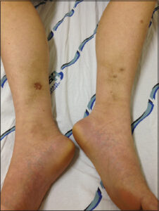
A twelve-year-old female patient sought dermatological treatment for brownish patches on her lower limbs. A skin biopsy revealed ochre dermatitis (Fig. 1). At fourteen years of age, the patient was sent to a vascular surgeon, who confirmed the diagnosis of ochre dermatitis. The patient was asymptomatic. Deep and superficial venous duplex scans were performed, which revealed no abnormalities in the venous system. Aminaphtone was prescribed, which led to the cessation of new patches and the continual fading of the existing patches until their complete disappearance. At 16, 19, and 22 years of age, the patient returned reporting that the brownish patches returned and also complained of social discomfort due to the unpleasant esthetic appearance of the hyperpigmentation. Aminaphtone was prescribed the second time and control of the patches was achieved. This study received approval from the Human Research Ethics Committee of the São José do Rio Preto School of Medicine, SP, Brazil #3.764.416.
DISCUSSION
The paper reports control of ochre dermatitis in an adolescent and the long-term evolution of the treatment. Ochre dermatitis is associated with chronic venous hypertension, yet there is a report of an association with probable capillary fragility [3]. The most striking occurrence in the present case was the emergence of ochre dermatitis in a patient beginning at twenty-two years of age.
The patient began treatment with a dermatologist, yet without a satisfactory result, and at twelve years of age, was sent to the vascular surgery service of the university. During the initial clinical evaluation, the occurrence of ochre dermatitis was confirmed, along with some isolated telangiectasias, fitting C1 of the CEAP classification. Venous Doppler revealed no abnormalities in the superficial or deep venous system, discarding the possibility of chronic venous hypertension. This finding lent support to the hypothesis of capillary fragility as the cause of the initial purpura that progressed to hyperpigmentation.
Another aspect to consider in the present case is the more appropriate diagnosis between ochre dermatitis and stasis dermatitis. There was no venous hypertension in the present case to suggest stasis dermatitis. This is important because there are reports of ochre dermatitis in patients with and without evidence of chronic venous hypertension. Therefore, capillary fragility may be an aggravating factor in patients with chronic venous hypertension, and studies suggest that the presence of iron ions may be an aggravating agent of the inflammatory process.
With regard to the treatment of stasis dermatitis, there are some reports on therapeutic options, yet with no emphasis on the physiopathological hypothesis of capillary fragility. The use of aminaphtone has recently been described as a therapeutic option in cases of ochre dermatitis and small hemorrhages [6]. In the present study, hyperpigmentation was controlled with the use of aminaphtone for three to four years, followed by a recurrence, which suggests that yet treatment is not curative and only achieves temporary control. A further prescription of the drug enabled control of the new patches. Thus, aminaphtone was useful for treatment and may be administered again in cases of recurrence.
Aminaphtone was prescribed at a dose of 75 mg twice a day for two months, during which there was no emergence of new purpura, and there was a progressive reduction in pigmentation until complete elimination. Thus, there was a slow resolution of hyperpigmentation, with fading of 30% to 40% after two or three months, and a complete disappearance over time.
CONCLUSION
Ochre dermatitis may be associated with capillary fragility, and the use of aminaphtone is a therapeutic option in such cases.
Consent
The examination of the patient was conducted according to the principles of the Declaration of Helsinki.
The authors certify that they have obtained all appropriate patient consent forms, in which the patients gave their consent for images and other clinical information to be included in the journal. The patients understand that their names and initials will not be published and due effort will be made to conceal their identity, but that anonymity cannot be guaranteed.
REFERENCES
1. Bergan J. Molecular mechanisms in chronic venous insufficiency. Ann Vasc Surg. 2007;21:260-6.
2. Sundaresan S, Migden MR, Silapunt S. Stasis dermatitis:Pathophysiology, evaluation, and management. Am J Clin Dermatol. 2017;18:383-90.
3. de Godoy. Treatment of stasis dermatitis using aminaphtone:A case series. J Med Case Rep. 2010;4:295.
4. Choonhakarn C, Chaowattanapanit S, Julanon N. Lipodermatosclerosis:A clinicopathologic correlation. Int J Dermatol. 2016;55:303-8.
5. Andraska EA, Horne DC, Campbell DN, Eliason JL, Wakefield TW, Coleman DM. Patterns of pediatric venous disease. J Vasc Surg Venous Lymphat Disord. 2016;4:422-5.
6. Pereira de Godoy JM, Macedo Paizan ML. Aminaphtone in the control of gingival bleeding in children. Drug Des Devel Ther. 2014;8:1331-4.
Notes
Request permissions
If you wish to reuse any or all of this article please use the e-mail (brzezoo77@yahoo.com) to contact with publisher.
| Related Articles | Search Authors in |
|
|
 http://orcid.org/0000-0002-1036-8779http://orcid.org/0000-0003-0425-4641http://orcid.org/0000-0001-9463-7608http://orcid.org/0000-0001-5424-7787 http://orcid.org/0000-0002-1036-8779http://orcid.org/0000-0003-0425-4641http://orcid.org/0000-0001-9463-7608http://orcid.org/0000-0001-5424-7787 |

Comments are closed.