Rapidly fatal metastatic melanoma arising from a congenital nevus in a young female
Jihad Kassel 1, Sara Elloudi1, Kaoutar Soussy2, Soukaina Chhiti1, Hanane Baybay1, Zakia Douhi1, Touria Bouhafa2, Fatima-Zahra Mernissi1
1Department of Dermatology, University Hospital Hassan II, Fes, Morocco, 2Radiation oncology department, Oncology hospital, University Hospital Hassan II, Fes, Morocco
Citation tools:


Copyright information
© Our Dermatology Online 2023. No commercial re-use. See rights and permissions. Published by Our Dermatology Online.
Sir,
Melanoma is an aggressive and potentially fatal tumor of melanocytic origin. It may occur at any age yet more rarely at a young age [1]. Melanomas in young patients have overall a more favorable prognosis than in older. However, progression to the metastatic stage and the death of the patient are not exceptional [2]. One of the major risk factors for the development of melanoma in children and young adults is the congenital melanocytic nevus (CMN) [3]. The risk of malignant transformation of all congenital nevi ranges from 0.05% to 10.7%. The risk of malignant degeneration is correlated with size and location [4]. The size of the CMN above 40 cm as well as the presence of satellite nevi and the location in the trunk seem to increase the risk of developing MM. The role of surgical removal in inducing melanomas is controversial. In anatomopathology, melanomas arising from CMNs are usually located in the dermis and hypodermis, while melanocytic proliferation in a melanoma without a CMN starts in the epidermis [3]. Given the differences in the anatomical involvement of the disease, melanoma arising from congenital nevi may be considered a separate entity from the conventional case of melanoma and management may differ. Large excision may not be sufficient to remove all neoplastic cells from the nevi, and adjuvant aggressive systemic therapies may be essential to avoid a fatal outcome [4]. A recent study revealed that congenital nevi preferentially harbor NRAS mutations rather than BRAF mutations commonly seen in other types of nevi, indicating an altered molecular basis of nevogenesis in congenital nevi [3]. Herein, we report the case of a rapidly fatal metastatic melanoma in a young female arising from a congenital nevus of the trunk.
A young female 24 years of age presented with a pigmented, congenital, 5 cm lesion of the abdomen. The patient underwent surgical excision of the lesion without histological assessment. A painful angiomatous nodule appeared over the existing lesion evolving for the last year (Fig. 1). The evolution was then marked three months later by the appearance of numerous erythematous and angiomatous cutaneous and subcutaneous nodules disseminated over the entire body (Figs. 2a and 2b). We also noted inguinal and axillary bilateral lymph nodes associated with asthenia, dyspnea, headaches, and dizziness. A biopsy of the angiomatous nodule adjacent to the nevus was performed and showed a predominant dermal proliferation with some intraepidermal nests of atypical melanocytic cells with foci of tumor necrosis and ulceration. IHC showed a positive marking of HMB45 and Melan A. A total body CT scan was performed showing metastases of the brain, lungs, soft tissue, pancreas, and bone with peritoneal calcinosis. The patient was transferred to the oncology and radiotherapy department and deceased two weeks later.
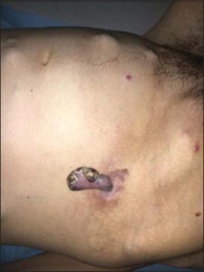 |
Figure 1: The pigmented angiomatous nodule above an asymmetric pigmented macule in the abdominal area. |
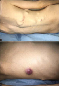 |
Figure 2: (a) Multiple subcutaneous nodules on the back. (b) The angiomatous nodule at the abdominal level. |
Consent
The examination of the patient was conducted according to the principles of the Declaration of Helsinki.
The authors certify that they have obtained all appropriate patient consent forms, in which the patients gave their consent for images and other clinical information to be included in the journal. The patients understand that their names and initials will not be published and due effort will be made to conceal their identity, but that anonymity cannot be guaranteed.
REFERENCES
1. Bebe FN, Hu S, Brown TL, Tulp OL. Metastatic melanoma in Florida, 1996-2010:Racial, demographic, occupational and tumor characteristics, and burden of metastasis. Our Dermatol Online. 2018;9:369-79.
2. Limam SAM, Erebih CE, Beyrouk A, Boye KI, Didi EH, Ely SO, et al. [Acral melanoma of the foot:A study of 9 cases and guidelines update]. Our Dermatol Online. 2019;10:23-9.
3. Lacoste C, Avril MF, Frassati-Biaggi A, Dupin N, Chrétien-Marquet B, MahéE, et al. Malignant melanoma arising in patients with a large congenital melanocytic naevus:Retrospective study of 10 cases with cytogenetic analysis. Acta Derm Venereol. 2015;95:686-90.
4. Wei CH, Shoo BA, Zedek DC, Kashani-Sabet M, Sagebiel RW, Leong SP. Rapidly lethal metastatic melanoma arising from a large congenital melanocytic naevus. BMJ Case Rep. 2009;2009:bcr09.2008.0981.
Notes
Request permissions
If you wish to reuse any or all of this article please use the e-mail (brzezoo77@yahoo.com) to contact with publisher.
| Related Articles | Search Authors in |
|
|
 http://orcid.org/0000-0002-5942-441Xhttp://orcid.org/0000-0003-3455-3810 http://orcid.org/0000-0002-5942-441Xhttp://orcid.org/0000-0003-3455-3810 |

Comments are closed.