Eccrine poroma on the forearm in a child: A rare presentation
Fatima Azzahra El Gaitibi 1, Hind Palamino1, Kaoutar Znati2, Laila Berbich1, Karima Senouci1
1Department of Dermatology, Mohammed V University in Rabat, Ibn Sina University Hospital, Rabat. Morocco, 2Department of Histopathology, Mohammed V University in Rabat, Ibn Sina University Hospital, Rabat. Morocco
Citation tools:


Copyright information
© Our Dermatology Online 2023. No commercial re-use. See rights and permissions. Published by Our Dermatology Online.
Eccrine poroma (EP) is a benign adnexal tumor arising from the terminal duct of the sweat gland. It represents 10% of all sweat gland tumors and is most commonly found on the sole or the side of the foot [1]. Herein, we report a case of EP on the right forearm in a child.
A thirteen-year-old female with no past medical history presented with an asymptomatic nodule on the right forearm (Fig. 1). The nodule had gradually increased in size and was not associated with pain, pruritus, or bleeding from the lesion. A physical examination revealed a firm, mobile flesh-colored nodule on the right forearm 7 × 6 mm in size. A dermoscopic examination revealed chalice-like vessels, whitish-pink areas, and yellow structureless areas (Fig. 2). The nodule was completely excised and histopathological findings showed a tumor proliferation connecting to the epidermis, organized on thick, cellular cords and composed of small, cohesive, round cells forming homogeneous layers. These cellular cords were separated by fibrous interstitial tissue, not especially inflammatory, and traversed by regularly distributed capillaries (Figs. 3 and 4). Based on these histological findings, a diagnosis of EP was reached.
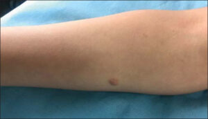 |
Figure 1: Flesh-colored nodule located on the right forearm. |
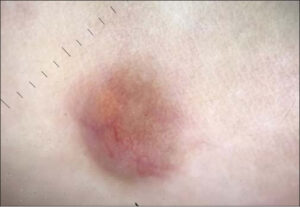 |
Figure 2: Dermoscopic image revealing chalice-like vessels, whitish-pink areas, and yellow structureless areas. |
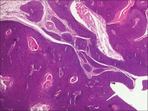 |
Figure 3: Histological image showing a tumor proliferation connecting to the epidermis, organized on thick, cellular cords separated by fibrous interstitial tissue (H&E, 20×). |
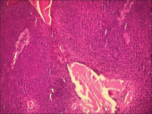 |
Figure 4: Histological image showing cellular cords composed of small, cohesive, round cells forming homogeneous layers (H&E, 40×). |
Although the pathogenesis of EP is unknown, it has been associated with trauma, scarring, and X-ray radiation [2]. EP occurs in middle-aged individuals, with only around ten cases reported in children. EP typically presents as a solitary, asymptomatic papule, nodule, or plaque gradually enlarging, with colors varying from flesh-colored to red, and brown, and bluish. The palm and sole are the most common localizations of this tumor. The main dermoscopic features are vascular structures, a white-to-pink halo surrounding vessels, pink-white structureless areas, and yellow structureless areas [2]. Pigmented EP may resemble basal cell carcinoma clinically and dermoscopically. Routinely, the diagnosis is reached by histopathology. Surgical excision is the treatment of choice. EP does not recur after excision. Malignant transformation rarely occurs [3].
Consent
The examination of the patient was conducted according to the principles of the Declaration of Helsinki.
The authors certify that they have obtained all appropriate patient consent forms, in which the patients gave their consent for images and other clinical information to be included in the journal. The patients understand that their names and initials will not be published and due effort will be made to conceal their identity, but that anonymity cannot be guaranteed.
REFERENCES
1. Ahmed jan N, Masood S. Poroma. 2020 Jul 8. In:StatPearls [Internet]. Treasure Island (FL):StatPearls Publishing;2021 Jan–.
2. Bombonato C, Piana S, Moscarella E, Lallas A, Argenziano G, Longo C. Pigmented eccrine poroma:Dermoscopic and confocal features. Dermatol Pract Concept. 2016;6:59-62.
3. Mantri MD, Dandale A, Dhurat RS, Ghate S. Pedunculated poroma on forearm:A rare clinical presentation. Indian Dermatol Online J. 2014;5:469-71.
Notes
Request permissions
If you wish to reuse any or all of this article please use the e-mail (brzezoo77@yahoo.com) to contact with publisher.
| Related Articles | Search Authors in |
|
|
 http://orcid.org/0000-0002-0313-6152 http://orcid.org/0000-0002-0313-6152 |

Comments are closed.