Apocrine hidrocystoma of the scalp: Additional case report
Misaki Kusano , Natuko Matumura, Tomoko Hiraiwa, Toshiyuki Yamamoto
Department of Dermatology, Fukushima Medical University, Fukushima 960-1295, Japan
Corresponding author: Dr. Misaki Kusano
Submission: 26.07.2020; Acceptance: 16.10.2020
DOI: 10.7241/ourd.20211.29
Cite this article: Kusano M, Matumura N, Hiraiwa T, Yamamoto T. Apocrine hidrocystoma of the scalp: Additional case report. Our Dermatol Online. 2021;12(1):94-95.
Citation tools:

Copyright information
© Our Dermatology Online 2021. No commercial re-use. See rights and permissions. Published by Our Dermatology Online.
Sir,
We have read with interest a recent report by Dahhouki et al. on a case of apocrine hidrocystoma in a rare location [1]. Herein, we describe an additional case of apocrine hidrocystoma that appeared on the scalp. The patient had a history of lung cancer, and a skin biopsy was performed to rule out skin metastasis.
An 80-year-old female, diagnosed with lung adenocarcinoma (pT1bN0M0, stage IA) two years before, was referred to our department complaining of an asymptomatic scalp nodule that appeared a few months prior to the referral. The lung cancer was surgically resected and the subsequent metastasis to the brain was treated with radiation. A physical examination revealed a smooth, dome-shaped, light blue nodule 1 cm in size on the scalp (Fig. 1). The patient had not received any treatment for the lesion. A skin biopsy was conducted to rule out skin metastasis. Histological features involved a cyst wall consisting of one to several layers of epithelial cells, some of which showing decapitation secretion indicative of apocrine secretion (Figs. 2a and 2b). The parietal cells did not show atypia and no inflammatory cell infiltration was observed. Immunohistochemistry demonstrated that the tumor cells were strongly positive for CEA and partially positive for 34 E12 (Figs. 3a and 3b), but negative for EMA, GCDFP-15, S-100, CK20, CAM5-2, and ……..-SMA. Therefore, a diagnosis of apocrine hidrocystoma was established.
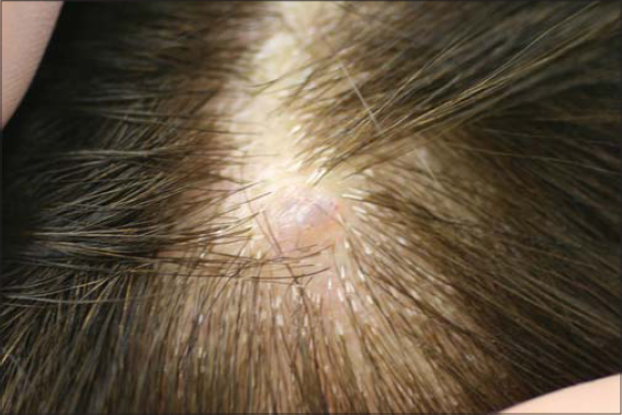 |
Figure 1: Smooth, dome-shaped, light blue nodule 5 mm in diameter on the scalp. |
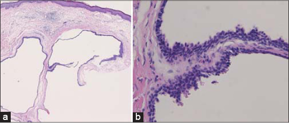 |
Figure 2: (a) Cystic structure in the dermis (H&E; 40×). (b) The epithelial cells with decapitation secretion further magnified (H&E; 200×). |
 |
Figure 3: The tumor cells (a) strongly positive for CEA and (b) partially positive for 34βE12 (H&E; (a-b): 200×). |
Apocrine hidrocystomas are benign cystic tumors that arise from the secretory portion of apocrine sweat glands in middle-aged and elderly people. They most frequently occur on the face—with a frequency of over 60%—followed by the trunk, extremities, and scalp [2]. According to an analysis of 167 Japanese cases conducted between 1968 and 2003 by Anzai et al. [3], tumors were most commonly found on the face (n = 102; 61.1%), followed by the trunk (23; 13.7%), the scalp (21; 12.6%), and the extremities (20; 12.0%). As far as we know, there have, since 2003, been three cases of apocrine hidrocystoma on the scalp [4–6]. One developed apocrine cystadenoma in a lesion associated with sebaceous nevus syndrome [4]. The other cases showed clinical features mimicking subcutaneous dermoid cysts [5] and a giant cell tumor [6]. We suggest that apocrine hidrocystoma ought to be considered as a differential diagnosis of cystic tumors on the scalp.
Consent
The examination of the patient was conducted according to the principles of the Declaration of Helsinki.
The authors certify that they have obtained all appropriate patient consent forms, in which the patients gave their consent for images and other clinical information to be included in the journal. The patients understand that their names and initials will not be published and due effort will be made to conceal their identity, but that anonymity cannot be guaranteed.
REFERENCES
1. Dahhouki S, Chaoui R, Kadiri SE, Douhi Z, Elloudi S, Baybay H, Mernissi FZ. Apocrine hidrocystoma of the scalp:Case report. Our Dermatol Online. 2020;11:e30.1-e30.2.
2. Magdaleno-Tapial J, Valenzuela-Oñate C, Martinez-Doménech Á, Marta García-Legaz-Martínez M, Martínez-Aparicio A, Alegre-de Miquel V, et al. Apocrine hidrocystoma on the nipple:The first report in this unusual location. Dermatol Online J. 2019;25:14.
3. Anzai S, Goto M, Fujiwara S, Da T. Apocrine hidrocystoma:A case report and analysis of 167 Japanese cases. Int J Dermatol. 2005;44:702-3.
4. Tejaswi C, Rangaraj M, Karthikeyan K. Apocrine hidrocystoma arising from nevus sebaceous on the scalp. Indian Dermatol Online J. 2016;7:111-3.
5. Azuma R, Yokokura H, Hiratsuka Y, Kakurai M, Umemoto N, Demitsu T. [A case of large apocrine hidrocystoma of the scalp with clinical features mimicking subcutaneous dermoid cyst]. Skin Surg. 2005;14:91-3.
6. Fujita H, Kai H, Yamamoto M, Mitomi H, Asahina A. Giant apocrine cystadenoma of the scalp. Eur J Dermatol. 2008;18:468-9.
Notes
Source of Support: Nil.
Conflict of Interest: None declared.
Request permissions
If you wish to reuse any or all of this article please use the e-mail (brzezoo77@yahoo.com) to contact with publisher.
| Related Articles | Search Authors in |
|
|
 http://orcid.org/0000-0003-0608-114X http://orcid.org/0000-0002-8390-2573 http://orcid.org/0000-0003-0608-114X http://orcid.org/0000-0002-8390-2573 |

Comments are closed.