Looking through the dermoscope in a case of piebaldism
Subrata Malakar1, Samipa Samir Mukherjee 2, Surit Malakar1
1Department of Dermatology, Rita Skin Foundation, Kolkata, India; 2Department of Dermatology, Cloud nine Hospital- Bangalore and Rita Skin Foundation, Kolkata, India
Corresponding author: Dr. Samipa Samir Mukherjee, E-mail: drsamipamukherjee@gmail.com
Submission: 20.06.2018; Acceptance: 25.03.2019
DOI: 10.7241/ourd.20193.28
Cite this article: Malakar S, Mukherjee SS, Malakar S. Looking through the dermoscope in a case of piebaldism. Our Dermatol Online. 2019;10(3):312-313.
Citation tools:
BibTex | CSV | RIS | refer/BiblX | Endnote XML | Wikipedia Citation Templates
Copyright information
© Our Dermatology Online 2019. No commercial re-use. See rights and permissions. Published by Our Dermatology Online.
Sir,
Piebaldism is an uncommon autosomal dominant disorder characterized by the congenital absence of melanocytes in localised areas of the skin and hair due to c-kit gene mutation, which affects the differentiation and migration of melanoblasts from the neural crest during the embryonic life. This condition is clinically characterised by the presence of localized stable hypo to depigmentation of the skin and hair, and by a characteristic distribution that involves the anterior trunk, extremities, the central portion of eyebrows, and the midfrontal portion of scalp with resultant white forelock, which is also the most common and persisting feature [1].
A 6 year old girl, first child born of non consanguineous marriage presented with asymptomatic white patches on the upper chest which remained stable since their appearance (Fig. 1). The child had no obvious ocular, hearing or neurological defects. The physical and mental development was normal. The audiogram of the child was normal. Routine biochemical tests were also normal. Family history was non contributory.
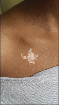
The examination revealed a well circumscribed white forelock in midfrontal region. Depigmented irregularly shaped macules were seen on the right upper chest. The entire back, abdomen, hands and feet, lower portions of forearm and legs and mucosae were completely spared. There was no evidence of involvement of eyebrows or eyelashes. The differentials considered at this point were piebaldism, nevus depigmentosus and congenital vitiligo.
Dermoscopic evaluation of the depigmented lesions revealed distorted melanocytic network with reduced pigment intensity(red arrows), absence of perifollicular pigment(green arrow), vast areas of depigmentation(yellow star) and islands of normal skin or hyperpigmentation (Figs. 2a and 2b).
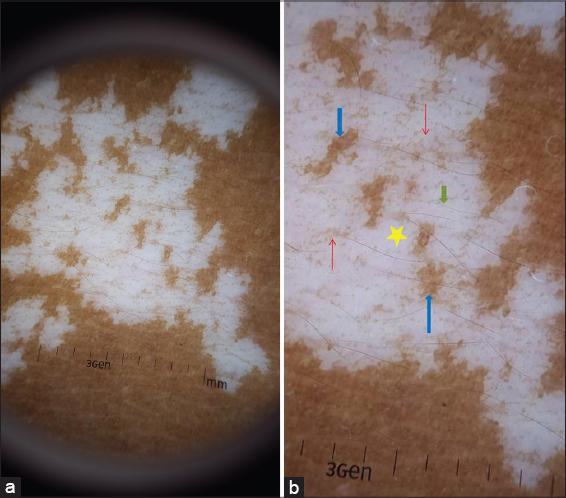
Based on clinical and dermoscopic features a diagnosis of piebaldism was established.
Piebaldism is a rare autosomal dominant disorder characterized by the congenital absence of melanocytes in affected areas of the skin and hair due to mutations of the c-kit gene, located on Chromosome 4q12, which affects the differentiation and migration of melanoblasts from the neural crest during the embryonic life [2]. Melanocytes are absent or considerably reduced in depigmented patches histologically and ultrastructurally. They are normal in number in the hyperpigmented areas.
Piebaldism results from defective migration of melanoblasts from neural crest to the ventral mid line thus manifesting dermoscopically as vast areas of depigmentation interspersed with islands of normal skin or hyperpigmentation. The second defect in the differentiation of melanoblasts to melanocytes results in distorted melanocytic network with reduced pigment intensity. We hypothesize that the absence of perifollicular pigment may be due to the defect in transfer of melanosomes from the melanocytes in the hair bud to the surrounding keratinocytes, however further studies and ultra structural analysis will be necessary to substantiate the same.
Herein we report the first ever description of dermoscopic features in a case of piebaldism and state that further continuous documentation of observations in these rare cases will help us in arriving at more specific features.
REFERENCES
1. Bassi A, Berti S, Galeone M. Piebaldism, QJM. An Int J Med. 2015;108:915.
2. Agarwal S, Ojha A. Piebaldism:A brief report and review of the literature. Indian Dermatol Online J. 2012;3:144-7.
Notes
Source of Support: Nil
Conflict of Interest: None declared.
Request permissions
If you wish to reuse any or all of this article please use the e-mail (brzezoo77@yahoo.com) to contact with publisher.
| Related Articles | Search Authors in |
|
|
|

Comments are closed.