|
Get Citation
|
|
|
El Jouari O, Gallouj S, Zinoune S, Baybay H, Mernissi FZ. Giant neurofi broma: a localization palpebral. Our Dermatol Online. 2018;9(3):329-330. |
|
|
Download citation file:
|
Giant neurofibroma: a localization palpebral
Ouiame El Jouari, Salim Gallouj, Safae Zinoune, Hanane Baybay, Fatima Zahra Mernissi
Department of Dermatology, Hassan II University Hospital, Fez, Morocco
Corresponding author: Dr. Ouiame EL Jouari, E-mail: eljouariouiame@gmail.com
DOI:10.7241/ourd.20183.26
Submission: 07.01.2018; Acceptance: 31.03.2018
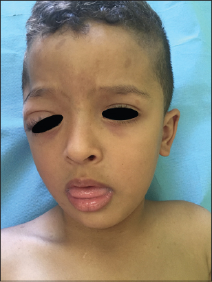
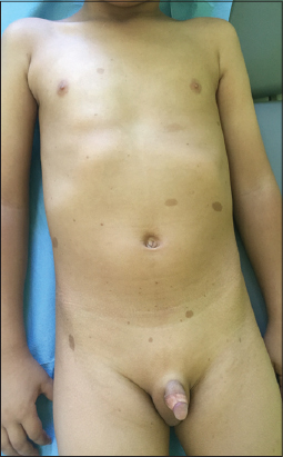
A 4-year-old child with asymptomatic upper right eyelid mass, observed by the mother since the age of one year, having progressively increased in size. A physical examination revealed a mass of 4 cm taking the upper right eyelid (Fig. 1), of soft consistency with ptosis (Fig. 2). Moreover, the child presented more than 6 coffee milk spots> 5 mm long axis, axillary lentigines without palpable adenopathies (Fig. 3). had confrmed that it was a plexiform neurofibroma associated with a dysplasty of the great right sphenoidal wing. the child was referred to the ophthalmology department for the management of his palpebral neurofibroma.

Neurofibroma is a manifestation of neurofibromatosis type 1 (NF1) or Von Recklinghausen disease, who is an oncogenic condition with an autosomal dominant inheritance pattern [1]. His incidence in children with NF1 is less than 10%. It is identified within the first few years of life. It follows the distribution of the trigeminal nerve [1]. It is manifested by a firm or soft palpebral mass with concomitant eyelid edema and it can lead to a ptosis or strabismus. Plexiform neurofibroma mostly occurs on the trunk and proximal extremities and presents as an occasionally pigmented, bag-like mass [2]. It is associated with pigmented spots (coffee coloured) in the skin, commonly seen on the back, abdomen and limbs (café au lait spots). Axillary freckling and lisch nodules may be present [3]. Magnetic resonance imaging (MRI) of the brain and orbits is needed to confirm diagnosis and to define its extent. The treatment is mainly surgical, it must be practiced early in order to avoid intraorbital extension and esthetic damage in children.
CONSENT
The examination of the patient was conducted according to the Declaration of Helsinki principles.
REFERENCES
1. Avery RA, Katowitz JA, Fisher MJ, Heidary G, Dombi E, Packer RJ, et al. Orbital/Periorbital Plexiform Neurofibromas in Children with Neurofibromatosis Type 1:Multidisciplinary Recommendations for Care. Ophthalmology. 2017;124:123-32.
2. Chang P, Meaux T, Calderon G. Solitary Neurofibroma. Our Dermatol Online. 2015;6:362-4.
3. Cukkemane A, Katheria V, Harinatha S. An interesting case of neurofibroma of face. Our Dermatol Online. 2016;7:479-81.
Notes
Source of Support: Nil
Conflict of Interest: None declared.
Comments are closed.