|
Get Citation
|
|
|
Oripelaye MM, Olasode OA, Onayemi O, Olanrewaju OF. Familial vitiligo in mother and child; the genetic theory connection. Our |
|
|
Download citation file:
|
Familial vitiligo in mother and child; the genetic theory connection
Muphy M. Oripelaye, Olayinka A. Olasode, Olaniyi Onayemi, Olatunde F. Olanrewaju
Department of Dermatology, Obafemi Awolowo University Teaching Hospitals Complex, Ile-Ife, Osun, Nigeria
Corresponding author: Dr. Muphy M. Oripelaye, E-mail: mmoripe@yahoo.co.uk
Submission: 11.09.2016; Acceptance: 03.02.2017
DOI: 10.7241/ourd.20172.46
ABSTRACT
Vitiligo is an acquired loss of skin pigment of unknown etiology. It frequently occurs in familial clusters thereby favoring the genetic theory for its pathogenesis. Several genes have been described in association with vitiligo and it is often thought to be polygenic with variable expressivity. We present two cases of familial vitiligo occurring in a mother and her child with a more severe presentation in the child. These cases of familial vitiligo portray the genetic theory with associated features suggesting genetic anticipation.
Key words: Vitiligo; Genetic anticipation; Genetic theory
INTRODUCTION
Vitiligo is an acquired loss of skin pigment of unknown etiology. Loss of melanocyte is demonstrated in the skin but the cause of loss of melanocyte is not known. Vitiligo affects about 0.4%-2%, of the general population [1]. The prevalence of vitiligo does not vary with gender nor race of affected patients [2]. Vitiligo poses a serious concern and reduces the quality of life because of increased contrast in the black skin and the myth associated with it [3].
One of the theories proposed to explain the etiology of vitiligo is the genetic theory with several genes described in association with vitiligo. We present a case of familial vitiligo in a mother and child with a probable tendency for genetic anticipation.
CASE REPORT
Case 1
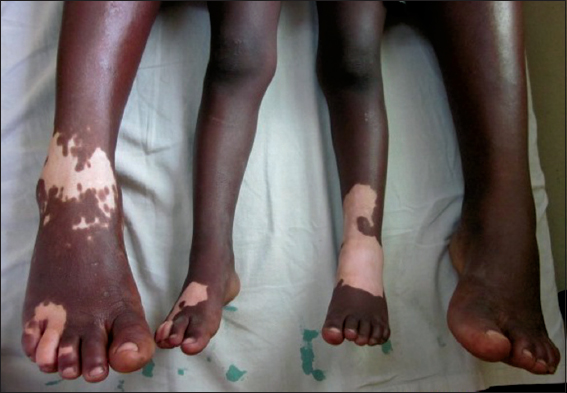
We report a 34-year-old Nigerian woman, who presented to our clinic on account of depigmented patches on the dorsum of the right foot. It started as a small macule and has increased progressively over 18 months to its present size (Figs. 1 and 2). There is no involvement of any other part of the body. There is no history suggesting any associated autoimmune disease. No history of emotional stress, or exposure to the industrial chemical in the patient. No other member of the family had vitiligo except the daughter. The results of laboratory investigations including complete blood count, blood sugar, and serum chemistry were within normal limit.

Case 2
The 3-years-old daughter of the patient (Case 1) developed depigmented patches on the dorsum of the foot about the same time with the mother. However, more areas were involved in the depigmentation including both feet (Figs. 2 and 3). No other part of the body was involved and there were no clinical findings suggesting autoimmune disease. The result of laboratory investigation were all normal.

Assessment of familial vitiligo was made and occurrence at the same time and in similar site in both the mother and the daughter was noted.
DISCUSSION
Familial vitiligo is frequently reported in the literature [4], and this has lent credence to the genetic theory of the pathogenesis of vitiligo. Transmissibility is thought to be polygenic with variable expressivity. The genes involved in the pathogenesis of vitiligo includes those associated with biosynthesis of melanin, response to oxidative stress and autoimmunity [5]. Genetically primed reduction in pro-opiomelanocortin peptides and α-MSH, as well as reduced expression of other genes involved in the melanocortin system (POMC, ASIP, MC1R, MC2R, MC3R, MC4R, MC5R) and melanogenesis (TRPI, DCT) has been described in association with vitiligo [6].
Bradley et al, in their work, suggested an association between catalase gene (CAT) and vitiligo [7]. Association between HLA system and vitiligo has been described with variable and inconsistent findings. Additionally, IFN-γ, TNF-α and IL-10 and their receptors have been shown to be associated with vitiligo [8].
A number of factors have been described in associated with the onset of vitiligo. These factors as shown by Behl et al include malnutrition, emotional trauma such as loss of Job, a death of a close relative, etc, and recurrent infections. Systemic antibiotic, topical chemical agent such as adhesive binds and wearing of rubber footwears were also associated with the onset of vitiligo. These factors were probably thought to be associated with vitiligo, by disturbing the immune balance and initiating autoimmune response [4].
The occurrence of vitiligo in the mother and her daughter as shown in our case strongly suggest familial predisposition. This may not be uncommon as it has been frequently reported in the literature [2]. However, the onset of vitiligo in the child at a younger age of 3 years in contrast to the mother in whom the onset was 34 years, and the increasing severity of the lesions in the child suggests a phenomenon akin to genetic anticipation. Genetic anticipation is a phenomenon whereby genetic diseases present earlier and with increased severity in succeeding generation. These are commonly reported in association with neurological diseases such as Huntington’s disease, myotonic dystrophy, spinocerebellar ataxia and Friedreich ataxia. It has also been demonstrated in association with Behçet’s disease by Fresko et al [9].
The occurrence of vitiligo lesions on the similar site (dorsum of the foot) was noted in the mother as well as the child. The similar location in both mother and child may actually be coincidental but it’s a food for thought. Environmental factors, as well as shared traits may have played a role.
While we consider the possibility of genetic anticipation, in these patients, we also gave consideration to the concurrent onset of the lesions in both the mother and the daughter. Since the onset of disease in both parent and offspring may not necessarily occur concurrently in genetic anticipation, we thought of the likelihood of two genetically predisposed individuals (mother and daughter) being exposed to triggers since they share the same environment. Although suspected, we did not identify any triggers in these patients. Several factors including malnutrition, emotional trauma, infections, antibiotics and topical chemical agents have described in association with the onset of vitiligo [4].
CONCLUSION
The cause of vitiligo remains unknown though familial predispositions being one of the proposed hypotheses for the etiology. While the genetic predisposition is thought to be polygenic, our cases also suggest the likelihood of genetic anticipation. We do recommend further study on the likelihood of genetic anticipation in familial vitiligo.
REFERENCES
1. Nanette BS. The epidemiology of vitiligo. Current Dermatol Report. 2015;4:36-43.
2. Kyriakis KP, Palamaras I, Tsele E, Michailides C, Terzoudi S. Case detection rates of vitiligo by gender and age. Int J Dermatol. 2009;48:328-9.
3. Linthorst Homan MW, Spuls PI, de Korte J, Bos JD, Sprangers MA, van der Veen JP. The burden of vitiligo: Patients characteristics associated with quality of life. J Am Acad Dermatol. 2009;61:411-20.
4. Behl P N, Agarwal A, Srivastava G. Etiopathogenesis of vitiligo: Are we dealing with an environmental disorder ? Indian J Dermatol Venereol Leprol [serial online]. 1999;65:161-7
5. Halder R, Taliaferro S. Vitiligo. Wolff K, Goldsmith L, Katz S, Gilchrest B, Paller A, Lefell D, eds. Fitzpatrick’s Dermatology in General Medicine. 7th ed. New York, NY: McGraw-Hill; 2008. Vol 1: 72.
6. Kingo K, Aunin E, Karelson M, Philips MA, Rästep R, Silm H, et al. Gene expression analysis of melanocortin system in vitiligo. J Dermatol Sci. 2007;48:113-22.
7. Casp CB, She JX, McCormack WT. Genetic association of the catalase gene (CAT) with vitiligo susceptibility. Pigment Cell Res. 2002;15:62-6.
8. Grimes PE, Morris R, Avaniss-Aghajani E, Soriano T, Meraz A. Topical tacrolimus therapy in vitiligo: Therapeutic responses and skin messenger RNA expression of proinflammatory cytokines. J Am Acad Dermatol. 2004;51:52-61.
9. Fresko I, Soy M, Hamuryudan V, Yurdakul S, Yavuz Ş, Tümerd Z, et al. Genetic anticipation in Behçet’s syndrome. Ann Rheum Dis. 1998:57:45-8.
Notes
Source of Support: Nil
Conflict of Interest: None declared.
Comments are closed.