Benign nevus with nerve sheath differentiation
Olfa El Amine El Hadj, Bouhaja Leila, Aida Goucha, Amor Gamoudi, Ahmed Elmay
Immuno-Histo-Cytological Department, Salah Azaiez Institute Tunis, Boulevard 9 Avril, Bab Saadoun 1006, Tunisia
Sir,
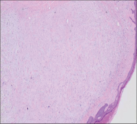
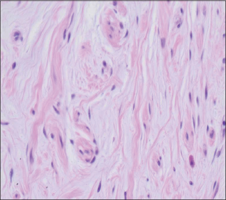
We report a 74 year-old woman who presented with lump on the right elbow. The decision was to remove the lump. The patient underwent a minor operation and the specimen was sent to the Histopathology department of Salah Azaiez institute. Grossly, an ellipse of skin was measuring 30 × 15mm with a central nodule measuring 10 × 8mm. It was transacted and 2 representative sections were put into blocks. Histological examination showed a well circumscribed dermal lesion made partly of nests of melanocytes (at the periphery of the lesion)and partly of spindle shaped cells with elongated and wavy nuclei (in the middle of the lesion), suggestive of a nerve sheath tumour (Fig. 1). There was no atypia and no mitoses (Fig. 2). The Immunohistochemical study showed that both the naevus cells and the spindled cells in the nerve sheat tumour-like area were positive for melaninA, S100 protein, EMA (Epithelial Membrane Antigen)and CD34. The desmin and SMA (smooth muscle actine)were negative. These features confirmed the diagnosis of benign nevus with nerve sheath differentiation. At 5 years of follow- up the patient was asymptomatic and there was no recurrence.
The patient’s informed consent was obtained.
Prior to the study, patient gave written consent to the examination and biopsy after having been informed about the procedure.
Neural differentiation by melanocytic nevi represents a well-recognized phenomenon, and melanocytic nevi with perineural differentiation have been reported [1]. Melanocytic nevi with neural differentiation are not rare. They retain some features of schwann cells and usually express S100 protein. Peripheral nerve sheath tumour and melanocytes are closely-related cells originating from the neural crest. It has been well-known that both benign and malignant melanocytic proliferation can show various type of neural differentiation [2,3]. Features of peripheral nerve sheath differentiation such as neuroid cords, nerve corpuscles, fascicle-like structures, and, exceptionally, palisading has been reported in melanocytic nevi [4]. Benign tumors of peripheral nerve sheath include mainly three subtypes: schwannoma, neurofibroma and perineurioma [5].
The main differential diagnoses for our case are benign naevus with nerve sheath differentiation or collision lesion tumour of naevus and nerve sheath tumour. The intimate mingling and merging of the naevus cells and the spindle-shaped cells suggest that this is a single lesion consisting of a benign intradermal naevus with nerve sheath differentiation. The immunohistochemistry results shows a mixture of S-100, EMA and CD34 positive cells.
Consent
The examination of the patient was conducted according to the Declaration of Helsinki principles.
Written informed consent was obtained from the patient for publication of this article.
REFERENCES
1. Wang L, Wang G, Gao T, Congenital melanocytic nevus with features of hybrid schwannoma/perineuriomaJ Cutan Pathol 2013; 40: 497-502.
2. Chen Y, Klonowski PW, Lind AC, Lu D, Differentiating Neurotized Melanocytic Nevi From Neurofibromas Using Melan-A (MART-1)ImmunohistochemicalStain Arch Pathol Lab Med 2012; 136: 810-5.
3. Hornick JL, Bundock EA, Fletcher CD, Hybrid schwannoma/perineurioma: clinicopathologic analysis of 42 distinctive benign nerve sheath tumorsAm J Surg Pathol 2009; 33: 1554-61.
4. Kroumpouzos G, Cohen LM, Intradermal melanocytic nevus with prominent schwannian differentiationAm J Dermatopathol 2002; 24: 39-42.
5. Weiss SW, Goldblum JR, Weiss SW, Goldblum JR, Benign tumors of peripheral nervesEnzinger and Weiss’s soft tissue tumors 2008; 5 ed. St. Louis, MO: Mosby Elsevier; 825-
Notes
Source of Support: Nil,
Conflict of Interest: None declared.
Comments are closed.