Clinical diagnosis and a short-term treatment of bullous pemphigoid in an adult Yemeni female: A case report
Mohamed A Al-Kamel
Regional Leishmaniasis Control Center (RLCC), P.O.Box 12692, Sana’a, Yemen
ABSTRACT
Bullous pemphigoid (BP) is an acquired, inflammatory, sub-epidermal, immunobullous disease. It usually occurs in the elderly population and reported cases of BP in adults are very rare. This report describes an adult Yemeni female who was clinically diagnosed to have BP, and successfully treated with a steroid containing short term regimen. It concludes the possibility of reaching a clinical diagnosis of BP in disadvantaged areas based on the clinical criteria, and the importance of considering a diagnosis of BP in young adults and even in children who present with itchy tense blisters of forearms, hands and feet. When accessible, further immunofluorescence studies are important to confirm the accuracy of the clinical diagnosis of BP.
Key words: Bullous; Pemphigoid; Adult; Clinical; Diagnosis; Treatment; Yemen
Abbreviations: BP, bullous pemphigoid; PV, bullous vulgaris; SJS, Stevens-Johnson syndrome; TEN, toxic epidermal necrolysis; SSSS, staphylococcal scalded skin syndrome; DH, dermatitis herpetiformis; EBA, epidermolysis bullosa acquisita; LABD, linear IgA bulous dermatosis; PCT, porphyria cutanea tarda; EM, erythema multiforme; NSAIDs, non-steroidal anti-inflammatory agents
INTRODUCTION
Bullous pemphigoid (BP) that was first described by Leverin 1953 is an acquired, acute or chronic, inflammatory, sub epidermal, immunobullous disease. Caused by autoantibody-mediated disruption of adhesion between basal keratocytes and the basement membrane [1,2].
Clinically, the earliest lesions may appear urticarial (like hives). Characteristic tense, usually symmetrical, ungrouped, with serous and/or hemorrhagic contents blisters of variable size eventually erupt either over normal or erythematous bases, most commonly at flexural areas of the limbs, trunk and abdomen. Any part of the skin surface can be involved; oral lesions have been reported, but other mucosal surfaces are usually not involved [2,3]. Clear diagnostic criteria can be lacking for definitive diagnosis in less than clear cut cases. If untreated, BP can persist for months or years, with periods of spontaneous remissions and exacerbations [4,5].
In Yemen, autoimmune disorders are among skin diseases groups with the lowest incidence rate. In general, BP is a rare, but the most commonly seen autoimmune blistering disease; most commonly occurs in elderly persons, but occasionally is seen in young adults and even in children [6,7].
This report describes an adult Yemeni female patient seen at Al Helal Hospital. Radaa district, of Al Baydaa governorate, in central Yemen, who was diagnosed to have BP based on history taking and physical examination, and successfully treated with a steroid-containing short-term regimen.
CASE REPORT
A 29-years-old Yemeni female was referred to my service in Nov. 27, 2015, with numerous itchy, tense vesicles and bullae on erythematous bases, located predominantly on her palms and soles, dorsa of hands and feet and ventral forearms (Fig. 1), with two crusted erosions on the lower lip and soft palate, of three weeks duration, insidious onset and progressive course. No ocular or genital lesions, and the trunk was spared.
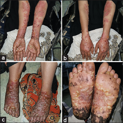
The blisters were sub-epidermal, filled either with clear or purulent fluid and concurrently occurred with diffuse pruritic urticarial patches which have started appearing few weeks prior to the visible blisters. The patient said that she evacuated some blisters by her hand.
Elicitation of clinical dermatological signs revealed positive Asboe-Hansen sign (Fig. 2), negative Nikolsky sign, and most of the solitary bullae showed regular rounded borders.

The patient reported a medical history with a frequent use of over-the-counter non-steroidal anti-inflammatory agents (NSAIDs), particularly ibuprofen to control a chronic daily headache. Prolonged sun-exposure was reported; past traumatic and family histories were unremarkable.
Review of systems reveals no other system complaints (fever, tachycardia, hypotension, altered level of consciousness, sore throat, chills, arthralgia, cough, dysuria, nor conjunctivitis).
Complete blood count, liver function and renal function tests were unremarkable, and the patients’ cardinal signs were unaffected. Biopsy for histology and immunofluorescence testing was unavailable. Thus, a provisional diagnosis of bullous pemphigoid (BP) of this patient was made based on the history and clinical examination.
She was hospitalized, and the treatment regimen included oral 60 mg/day prednisolone as a starting dose (gradually tapered over the next17 days), oral azithromycin 500 mg/day for 6 days, betamethasone-clioquinol containing topical cream (Betnovate-C™) thrice a day and pheniramine maleate injections (first-generation antihistamines) twice daily. Intravenous fluids, Vitamins A, E and C tablets and potassium permanganate as soaks (diluted as 1:1000) were prescribed as an adjuvant therapy.
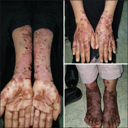
The patient left the hospital on the 2nd day upon her request. On the 7th day, she was presented for a follow-up with complete remission of the blisters, but with post-inflammatory erythematous patches (Fig. 3). The patient was advised for regular future follow-up visits.
DISCUSSION
The diagnosis of bullous pemphigoid (BP) is generally made based on the clinical features which may be seen in patients with all forms of BP which include significant pruritus which is frequently present weeks or months before the appearance of any visible skin lesion and may be the only manifestation of the disease and tense fluid filled sub-epidermal blisters that do not rupture easily, supplemented by histologic features. Palmoplantar involvement with vesiculobullous lesions may also represent the only feature of the disease (dyshidrosiform pemphigoid), or occur in association with other more typical widespread lesions. Localized forms of BP account for up to 30 percent of cases. Blisters involves the mucosa in up to a quarter of patients [8–11].
Other clinical dermatological signs such as Asboe-Hansen sign (+ve in BP) and Nikolsky sign (-ve in BP) gave a clue to the probable possible diagnosis of BP [12].
In the current case, I relied on these cardinal symptoms and clinical signs to reach a clinical diagnosis of BP at such a suburban area lacking the advanced diagnostic.
Several triggers have been anecdotally implicated in the development of BP, such as ultraviolet irradiation, X-ray therapy, and exposure to some drugs. In the current case, non-steroidal anti-inflammatory agents (NSAIAs) and prolonged sun exposure are suspected trigger factors.
This patient had some clinical events, suggesting other blistering disorders which were considered and then ruled out on the basis of at least one characteristic clinical feature that distinguish each of them from BP, as the following:
Positive Asboe-Hansen sign and negative Nikolsky’s helped in the exclusion of Stevens-Johnson syndrome (SJS), Hailey-Hailey disease, staphylococcal scalded skin syndrome (SSSS), and pemphigus vulgaris (PV); SJS and toxic epidermal necrolysis (TEN) are characterized by a prodrome of malaise and fever, and mucosal membranes are affected in 92 to 100 percent of patients, usually at two or more distinct sites (ocular, oral, and genital); TEN involves sloughing of greater than 30 percent of the body surface area; Skin lesions in PV are usually erosive, painful (but very rarely pruritic), superficial and do not form tense bullae; Blisters in dermatitis herpetiformis (DH) are the result of gluten sensitivity, often associated with other autoimmune disorders, and the lesions usually do not appear in the mouth; In epidermolysis bullosa acquisita (EBA), blisters tend to appear both spontaneously and as a result of trauma, predominantly on trauma-exposed body surfaces, and lesions heal with significant scarring; In linear IgA bulous dermatosis (LABD), blisters arise on normal, erythematous, or urticarial skin with a characteristic ‘string of pearls’ appearance; In porphyria cutanea tarda (PCT), patients present with blisters on sun-exposed areas of the body such as face, neck and hands, and the lesions are often painful, heal slowly with atrophic scars, milia, and post-inflammatory hyperpigmentation; In erythema multiforme (EM), the hallmark of which is the iris or target lesion [13–16].
As per this case, a short-term treatment regimen containing high dose of oral steroids, oral azithromycin, and potent topical steroids is an effective option in the treatment of BP.
CONCLUSION
This report concludes the possibility of reaching a clinical diagnosis of BP in disadvantaged areas through careful clinical examination, and the importance of considering a diagnosis of BP in young adults and even in children who present with itchy tense blisters of forearms, hands and feet. When accessible, further immunofluorescence studies are important to confirm the accuracy of the clinical diagnosis of BP.
Consent
The examination of the patient was conducted according to the Declaration of Helsinki principles.
REFERENCES
1. Lever WF, PemphigusMedicine (Baltimore) 1953; 32: 1-123.
2. Wolff K, Goldsmith L, Gilchrest B, Wolff K, Goldsmith L.A, Katz S, Stephen. “Chapter 54. Bullous Pemphigoid”Fitzpatrick’s Dermatology In General Medicine, Two Volumes 2007; 7th ed. Mcgraw-hill;
3. Di Zenzo G, Della Torre R, Zambruno G, Borradori L, Bullous pemphigoid: From the clinic to the benchClin Dermatol 2012; 30: 3-16.
4. Lipsker D, Borradori L, Bullous pemphigoid: Whatare you? Urgent need of definitions and diagnostic criteriaDermatology 2010; 221: 131-4.
5. Agrawal J, Prashanth SK, Chandra J, Veena KM, Chatra L, Drug-Induced Bullous Pemphigoid: Expect the UnexpectedWorld J Dentistry 2011; 2: 151-3.
6. Al-Kamel MA, Spectrum of winter dermatoses in rural YemenInt J Dermatol 2015; Sep4
7. Ujiie H, Shibaki A, Nishie W, Shimizu H, What’s new in bullous pemphigoidJ Dermatol 2010; 37: 194-204.
8. Swagata T, Stefanie H, Luca B, Clinical Challenges and Recent Advances in the Diagnosis of Bullous PemphigoidExpert Rev Dermatol 2013; 8: 407-16.
9. Tran JT, Mutasim DF, Localized bullous pemphigoid: a commonly delayed diagnosisInt J Dermatol 2005; 44: 942-
10. Khandpur S, Verma P, Bullous pemphigoidIndian J Dermatol Venereol Leprol 2011; 77: 450-5.
11. Bakker CV, Terra JB, Pas HH, Jonkman MF, Bullous pemphigoid as pruritus in the elderly: a common presentationJAMA Dermatol 2013; 149: 950-3.
12. Sentamilselvi G, Eponymous Dermatological Signs in Bullous DermatosesIndian J Dermatol 2014; 59: 21-3.
13. Letko E, Papaliodis DN, Papaliodis GN, Daoud YJ, Ahmed AR, Foster CS, Stevens-Johnson syndrome and toxic epidermal necrolysis: a review of the literatureAnn Allergy Asthma Immunol 2005; 94: 419-
14. Bastuji-Garin S, Rzany B, Stern RS, Shear NH, Naldi L, Roujeau JC, Clinical classification of cases of toxic epidermal necrolysis, Stevens-Johnson syndrome, and erythema multiformeArch Dermatol 1993; 129: 92-
15. Bystryn JC, Rudolph JL, PemphigusLancet 2005; 366: 61-73.
16. Bronwyn S, Fauzia R, Amor K, Bullous Pemphigoid: A Short ReviewDermatol Nursing 2009; 21: 322-6.
Notes
Source of Support: Nil,
Conflict of Interest: None declared.
Comments are closed.