Cutaneous creeping eruption in a child
Shrikiran Aroor, Sandeep Kumar, Suneel Mundkur
Department of Pediatrics, Kasturba Medical College, Manipal University, Manipal, India
Sir,
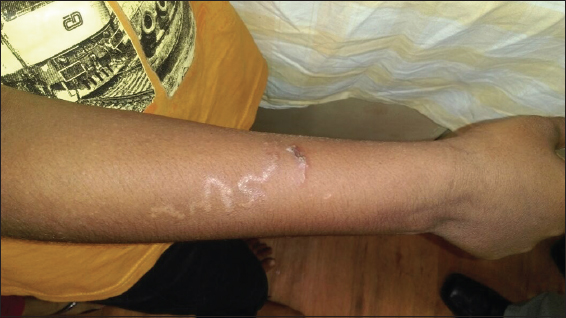
A 6 year-old boy from coastal area presented with history of intensely pruritic skin lesions over the right forearm for last 3 days. There were no other symptoms. The boy used to play on the beach barefoot daily. Clinical examination revealed an erythematous, serpentine lesion on the dorsal aspect of the right forearm (Fig. 1). Systemic examination was unremarkable. Hemogram was normal except for eosinophilia (absolute eosinophil count-1400/mm3). A diagnosis of cutaneous larva migrans was made and he was treated with single dose of albendazole (400 mg) and ivermectin (6 mg). Lesion had healed during his subsequent followup after 1 week.
Cutaneous larva migrans (CLM) also called creeping eruption, plumber’s itch, is characterized by classical serpentine skin lesions in a tropical setting [1]. Bare foot walkers, farmers, children playing in beaches, sandy and moist arears are at high risk. CLM is mainly caused by infection with larvae of animal hookworms like Ankylostoma caninum and A. braziliens. Other offenders include A. ceylonicum,Bubostomum phlebotomum etc [2,3]. Larval penetration of skin and migration cause itchy erythematous, raised vesicular and serpentine cutaneous lesion. The disease is usually self-limiting and lasts for 4-6 weeks until the larva dies and humans are accidental and ‘dead-end’ host [2,4]. Severe infestations manifest as Loeffler’s syndrome of pulmonary eosinophilia and rarely as eosinophilic enteritis [5]. Biopsy is of no value as the larvae advance ahead of the clinical tract. Optical coherence tomography is a non-invasive modality for diagnosis [6]. We treated with a single dose of ivermectin and albendazole [7]. Other treatment regimens include oral albendazole (400 mg) daily for 3 days and topical application of 10% thiabendazole [8]. CLM can be prevented by avoiding skin contact with soil contaminated with animal feces and adequately covering the feet when visiting sandy and moist areas.
Consent
The examination of the patient was conducted according to the Declaration of Helsinki principles.
REFERENCES
1. Yap FB, Creeping eruptionInt J Infect Dis 2010; 14: e545.
2. Brenner MA, Patel MB, Cutaneous larva migrans: the creeping eruptionCutis 2003; 72: 111-5.
3. Purdy KS, Langley RG, Webb AN, Walsh N, Haldane D, Cutaneous larva migransLancet 2011; 377: 1948.
4. Criado PR, Belda W, JrVasconcellos C, Silva CS, Cutaneous larva migrans: a bad souvenir from the vacationDermatol Online J 2012; 18: 11.
5. Butland RJ, Coulson IH, Pulmonary eosinophilia associated with cutaneous larva migransThorax 1985; 40: 76-7.
6. Morsy H, Mogensen M, Thomsen J, Thrane L, Andersen PE, Jemec GB, Imaging of cutaneous larva migrans by optical coherence tomographyTravel Med Infect Dis 2007; 5: 243-6.
7. Coumas E, Datry A, Paris L, Efficacy of ivermectin in the treatment of cutaneous larva migransArch Dermatol 1992; 128: 994-5.
8. Chiriac A, Birsan C, Chiriac AE, Murgu A, Solovan C, Cutaneous larva migrans: Report of three cases with excellent response to albendazoleOur Dermatol Online 2012; 3: 126-7.
Notes
Source of Support: Nil,
Conflict of Interest: None declared.
Comments are closed.