A rare presentation of an ectopic breast tissue in axilla
Radhika Vidyasagar, P. Sudarshan, N. Ravindranath Singh, S. Shivaram
Department of General Surgery, MVJ Medical College, Bangalore, India
ABSTRACT
Accessory breast tissue is rare accounting to less than 1% cases seen in females. It is usually bilateral. We report a case of 24-year-old woman with a lump in the left axilla in view of its rarity and made a differential diagnosis of fibroadenoma, which following the investigations and histopathological report was confirmed as revealed fibroadenoma in the axilla. It should also be considered as a differential diagnosis for all axillary swellings.
Key words: Axilla; Fibroadenoma; Breast
INTRODUCTION
Accessory breast tissue is commonly located above the breast in the axilla. It is uncommon and usually bilateral. Accessory breast tissue may be seen as an enlarging mass in the axilla during pregnancy and persists, as excess tissue in the axilla after lactation is complete. The accessory mammary tissue may be removed surgically if it is large or cosmetically deforming or to prevent enlargement during future pregnancy.
We report a case of 24-year-old woman with a lump in the left axilla in view of its rarity that was histologically identical to the fibro adenoma seen in the breast and also to consider for differential diagnosis of axillary mass.
CASE PRESENTATION
24-year-old woman presented with lump in the axilla since 2 months that increased in size and associated with discomfort. Patient has no family history of breast cancer.
On examination, there was a lump in the left axilla measuring 3 × 2 cms that was firm in consistency, mobile, non-tender. Skin over the swelling was normal. There were no nipple or areola changes seen. Both the breast and right axilla were clinically normal. A provisional differential diagnosis of Axillary lymphadenopathy was made.
Prior to the study, patient gave written consent to the examination and biopsy after having been informed about the procedure.
INVESTIGATIONS
Mammography of the right breast is normal. The visualized left axillary region reveals a focal bulge of homogenous soft tissue density of size approximately 2 × 2 cm, suggestive of benign SOL in axilla fibroadenoma.
Fine needle aspiration cytology (FNAC) report done outside and in our hospital showed breast tissue with an encapsulated tumor composed of glands and stroma. The glands are predominantly in a pericanalicular pattern, some areas show intracanalicular arrangement. The glands are lined by double-layered epithelium surrounded by proliferative stroma suggestive of fibroadenoma.
TREATMENT
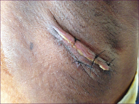
Patient underwent excision biopsy on 5/6/2014. Histopathological examination revealed a globular, glistening soft tissue mass and the cut section showed pale white with slit like spaces (Figs. 1 and 2).

DISCUSSION
Polymastia is a term used to describe the presence of more than 2 breasts in human beings. It is synonymous with supernumerary or accessory breast tissue [1]. Accessory breast tissue has the potential to undergo the same benign and malignant changes as normal pectoral breast tissue.
The differential diagnosis for a solitary axillary mass may range from breast parenchymal lesions, lymph nodes, and infectious and vascular lesions, as well as an axillary tail of spence (extension of normal parenchyma towards the axilla) or a torn muscle belly. It is important to differentiate benign from malignant axillary masses to avoid unnecessary concern and intervention.
During the 6th week of embryonic development, the mammary milk lines, which represent 2 ectodermal thickenings, develop along the sides of the embryo, extending from the axillary region to the groin [1]. In normal development, most of the embryologic mammary ridges resolve, except for 2 segments in the pectoral region, which later become breasts.
Failure of any portion of the mammary ridge to involute can lead to ectopic breast tissue with (polythelia) or without (polymastia) a nipple-areolar complex. Therefore, ectopic breast usually occurs along the “milk line” or mammary line [2].
In 1915, Kajava published a classification system for supernumerary breast tissue that remains in use today. Class I consists of a complete breast with nipple, areola, and glandular tissue. Class II consists of nipple and glandular tissue but no areola. Class III consists of areola and glandular tissue but no nipple. Class IV consists of glandular tissue only. Class V consists of nipple and areola but no glandular tissue (pseudomamma). Class VI consists of a nipple only (polythelia) [1]. Class VII consists of an areola only (polythelia areolaris). Class VIII consists of a patch of hair only (polythelia pilosa). The accessory tissue may range from a subcutaneous focus of breast tissue to a full accessory breast complete with areola and nipple. The presence of a small nipple is the most frequently noted accessory breast structure.
The clinical significance of the polymastia lies in the fact that apart from the psychological and cosmetic impact, it develops the same pathological changes as the normally located breast tissue such as inflammation, fibrosis, fibroadenoma, cystosarcoma phyllodes, and carcinoma [3]. Diagnosis of EBT is strongly suggested by history of cyclic changes during the menstrual period or by initial appearance during pregnancy. Pathology in EBT should be evaluated by the same methods as in normal breast tissue. Radiological examination should be done to rule out the urogenital malformation as supernumerary kidneys, renal agenesis, and carcinomas [4].
Misdiagnosis of accessory breast tissue is common and the most common presumptive diagnoses include lipoma, lymphadenopathy, hidradenitis, sebaceous cyst, vascular malformation, and malignancy.
On mammography, accessory breast tissue in the axilla resembles normal glandular parenchyma and is separate from the breast, unlike the axillary tail of Spence, which is a normal direct extension toward the axilla from the main breast tissue.
The treatment of choice for symptomatic accessory axillary breast tissue is surgical excision. Cosmesis is the main indication in the majority of cases [5].
Removal of the tissue will relieve the physical discomfort and also confirms the diagnosis.
CONCLUSION
Fibroadenoma in the ectopic breast tissue is indeed a rare occurrence. It should also be considered as a differential diagnosis for all axillary swellings.
Consent
The examination of the patient was conducted according to the Declaration of Helsinki principles.
REFERENCES
1. Amaranathan A, Balaguruswamy K, Bhat RV, Bora MK, An ectopic breast tissue presenting with fibroadenoma in axillaCase Rep Surg 2013; 2013: 947295.
2. Shin SJ, Sheikh FS, Allenby PA, Rosen PP, Invasive secretory (juvenile) carcinoma arising in ectopic breast tissue of the axillaArch Pathol Lab Med 2001; 1251372-4.
3. Rizvi G, Pandey H, Gupta MK, Fibroadenoma of ectopic breast tissue in axillaJ Case Reports 2012; 213-15.
4. Jethwani U, Saroha R, Verma R, Fibroadenoma of ectopic breast of axilla: A rare case reportClin Canc Invest J 2015; 4: 102-4.
5. Lesavoy MA, Gomez-Garcia A, Nejdl R, Yospur G, Syiau TJ, Chang P, Axillary breast tissue: clinical presentation and surgical treatmentAnn Plast Surg 1995; 35: 356-60.
Notes
Source of Support: Nil
Conflict of Interest: None declared.
Comments are closed.