Embolis cutis medicamentosa, a rare preventable iatrogenic complication
Kalegowda Deepadarshan, Manjunath Kavya, Muddanahalli Rajegowda Harish, Basavapura Madegowda Shashikumar
Department of Dermatology, Mandya Institute of Medical Sciences, Mandya-571401, Karnataka, India
ABSTRACT
Embolis cutis medicamentosa is an uncommon iatrogenic complication characterised by variable degree of skin and tissue necrosis, likely to follow intramuscular injection. Intense pain and purplish discoloration of overlying skin, with or without reticulate pattern subsequently followed by tissue necrosis and scarring is highly specific for this syndrome. It has also been reported following intravenous, intra-articular and subcutaneous injections. Herein we are reporting two cases of this rare preventable entity.
Key words: Embolia cutis medicamentosa; tissue necrosis; intramuscular; diclofenac
INTRODUCTION
Embolia cutis medicamentosa also known as Nicolau syndrome, livedoid dermatitis characterised by severe painful local necrosis at the site of injected medicament [1,2]. Intense pain and purplish discoloration of overlying skin, with or without reticulate pattern subsequently followed by tissue necrosis and scarring is highly specific for this syndrome. Initially described by Nicolau and Frudenthal as an adverse effect with use of bismuth salts intramuscularly in syphilis patients [3]. Clinically, patient manifests with intense pain at the injection site, followed by bluish discoloration of overlying skin assuming reticulate pattern, referred as non-inflammatory retiform purpura, livedo-like dermatitis.2 In later stages, discoloured area undergoes necrosis and ulceration may involve sub-cutis and underlying muscles.
CASE REPORTS
Case 1
A 50 year old male presented with non healing ulcer over the left buttock of one month duration. Patient had fever and generalized body ache one month back, for which he was administered intramuscular injection of diclofenac in outer aspect of left buttock. Immediately after the injection, patient experienced intense pain along with redness followed by bluish discoloration of the overlying skin after two days. Later the entire skin became black with necrosis which eroded over a week. On examination, a solitary well defined ulcer measuring 10×5 cm in size, covered with necrotic tissue (Fig. 1) was noted over the outer aspect of left buttock. Patient was treated with oral and topical antibiotics, without much improvement. Patient was admitted, wound was debrided, and was started on systemic antibiotics. Routine investigations including bleeding time, clotting time were normal. Following daily dressings and systemic antibiotics, wound was covered with healthy granulation tissue in about 2 weeks. Split skin grafting was done to hasten the healing.
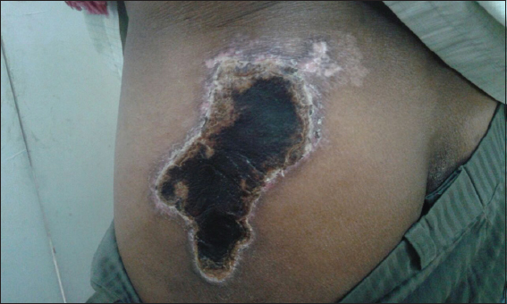
Figure 1: Case 1 showing well defined ulcer measuring 10×5 cm in size, covered with necrotic tissue.
Case 2
A 45 year old female presented with chronic non-healing ulcer of one month duration over the outer aspect of right buttock. Patient had received intramuscular injection of diclofenac for joint pain 3weeks back. Immediately following injection she developed intense pain and blanching of skin. Later over a period of 2 weeks entire blanched area becomes ulcerated and necrotic. On examination, a solitary well defined ulcer measuring 4×6 cm in size, localized to outer aspect of right buttock covered with necrotic tissue was noted (Fig. 2). Patient was admitted and wound debridement was done. After daily dressings and antibiotics, ulcer showed signs of healing. Finally after 2 weeks it healed with scar formation.
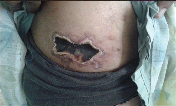
Figure 2: Case 2 showing well defined ulcer localized to outer aspect of right buttock covered with necrotic tissue.
DISCUSSION
Embolia cutis medicamentosa or Livedoid-like dermatitis (Nicolau syndrome) is an uncommon iatrogenic complication characterised by variable degree of skin and tissue necrosis, likely to follow intramuscular injection [2]. Cases have been reported following intramuscular injection of vitamin-K [4], diclofenac [5], bismuth [6], benzathine penicillin [7], gentamycin [8] and vaccination [9]. It has also been reported following intravenous polidocanol 1% [10], intra-articular corticosteroids [11], subcutaneous injections of pegylated interferon-α [12] and sclerotherapy for pyogenic granuloma [13].
The exact etio-pathogenesis of this syndrome is not known. Probable explanations for the ischemic necrosis of the skin and deep tissues seen in this syndrome includes, vasospasm secondary to needle prick, pressure of material placed around vessel and embolization of the injected material [7]. Cutaneous necrosis seen following bismuth salt injections in syphilis patients were thought to be due to high viscosity of salts causing blockage arterioles. Application of cold compresses can aggravate necrosis [14].
Complications like paralysis of limb has been reported which is attributed to embolization of medication into the internal iliac vessels [15]. Late complications such as contractures and deformities can occur due to scarring process that needs surgical correction [6]. Soft tissue sarcoma can occur as a rare complication at the site of tissue necrosis [16].
Diagnosis is mainly clinical. Nicolau syndrome has to be differentiated from necrotising fasciitis. In the latter, there is rapidly spreading infection of the sub-cutis and fascia due to haemolytic streptococci and other anaerobes. Necrotising fasciitis usually occurs following surgery or after a deep penetrating injury and manifests with intense pain with erythema of the skin later becomes purplish and ulcerates. On X-ray there is often presence of air in the tissues. Ultrasound and magnetic resonance imaging often helpful to delineate the extent of damage in the tissues [2].
Early institution of treatment has been reported to avert the extent of necrosis. Measures to improve the vascularity such as pentoxyphylline, hyperbaric oxygen, intravenous alprostadil and thrombolysis with heparin has been tried in the immediate post-event period [2]. Surgical debridement and systemic antibiotics play pivotal role in controlling infection. Since it can occur to common drugs it is very important for the Dermatologists to be aware of this rare iatrogenic disfiguring condition.
CONSENT
The examination of the patient was conducted according to the Declaration of Helsinki principles.
REFERENCES
1. Breathnach SM, Burns T, Breathnach S, Cox N, Griffiths C, Drug reactionsRooks Textbook of Dermatology 2010; 8th ed. Oxford: Blackwell Publishing; 75.1-160.
2. Nischal K, Basavaraj H, Swaroop M, Agrawal D, Sathyanarayana B, Umashankar N, Nicolau Syndrome: An Iatrogenic Cutaneous NecrosisJ Cutan Aesthet Surg 2009; 2: 92-5.
3. Kohler LD, Schwedler S, Worret WI, Embolia cutis medicamentosaInt J Dermatol 1997; 36: 197
4. Koklu E, Sarici SU, Altun D, Erdeve O, Nicolau syndrome induced by intramuscular vitamin K in a premature newbornEur J Pediatr 2009; 168: 1541-2.
5. Murthy SC, Siddalingappa K, Suresh T, Nicolau’s syndrome following diclofenac administration: A report of two casesIndian J Dermatol Venereol Leprol 2007; 73: 429-31.
6. Corazza M, Capozzi O, Virgilit A, Five cases of livedo-like dermatitis (Nicolau’s syndrome) due to bismuth salts and various other non-steroidal anti-inflammatory drugsJ Eur Acad Dermatol Venereol 2001; 15: 585-8.
7. De Sousa R, Dang A, Rataboli PV, Nicolau syndrome following intramuscular benzathine penicillinJ Postgrad Med 2008; 54: 332-4.
8. Kim DH, Ahn HH, Kye YC, Choi JE, Nicolau syndrome involving whole ipsilateral limb induced by intramuscular administration of gentamycinIndian J Dermatol Venereol Leprol 2014; 80: 96
9. Kienast AK, Mentze D, Hoeger PH, Nicolau’s syndrome induced by intramuscular vaccinations in children: Report of seven patients and review of the literatureClin Exp Dermatol 2008; 33: 555-8.
10. Geukens J, Rabe E, Bieber T, Embolia cutis medicamentosa of the foot after sclerotherapyEur J Dermatol 1999; 9: 132-3.
11. Cherasse A, Kahn MF, Mistrih R, Maillard H, Strauss J, Tavernier C, Nicolau’s syndrome after local glucocorticoid injectionJoint Bone Spine 2003; 70: 390-2.
12. Sonntag M, Hodzic-Avdagic N, Bruch-Gerharz D, Neumann NJ, Embolia cutis medicamentosa after subcutaneous injection of pegylated interferon-αHautarzt 2005; 56: 968-9.
13. Nirmal B, Segu SS, Sacchidanand SA, Deshpande P, Nicolau syndrome following sclerotherapy for pyogenic granulomaIndian J Dermatol Venereol Leprol 2014; 80: 484
14. Senel E, Ada S, Güleç AT, Ca?lar B, Nicolau syndrome aggravated by cold application after i.m. diclofenacJ Dermatol 2008; 35: 18-20.
15. Miranda MC, Rozenfeld S, Olivera SP, A systematic review of the nonallergic adverse reactions following benzathine penicilline injectionsJ Vasc Br 2004; 3: 253-60.
16. Mayrink M, Mendonça AC, da Costa PR, Soft-tissue sarcoma arising from a tissue necrosis caused by an intramuscular injection of diclofenacPlast Reconstr Surg 2003; 112: 1970-1.
Notes
Source of Support: Nil,
Conflict of Interest: None declared.
Comments are closed.