A unique case of tiny disseminated angiomas from childhood: a variant of “petechial” angiomata?
Yuka Hanami, Toshiyuki Yamamoto, Mikio Masuzawa
Department of Dermatology, Fukushima Medical University, Fukushima, Japan
Sir,
A 14-year-old girl was referred to our department complaining of a number of angiomas on the extremities. The patient stated that the angiomas had appeared on the upper limbs at the age of 10 and had gradually increased in number. Physical examination revealed a number of pin-sized to 2-mm-diameter reddish angiomas on the outer side of the upper arms, forearms, and thighs. She refused to undergo a skin biopsy. Three years later, she visited us again because of a further increase in the number of the angiomas. On physical examination, a number of small reddish nodules were scattered on the thighs, back, upper limbs, and abdomen (Figs. 1–3). Telangiectasia was also observed on the upper arm and chest (Fig. 4). Results of laboratory examinations including liver and renal function were normal. Other findings, suggestive of POEMS (Polyneuropathy, Organomegaly, Endocrinopathy, Monoclonal protein, and Skin changes) syndrome or Fabry disease, were not accompanied. Unfortunately, the patient once again refused examination by skin biopsy.

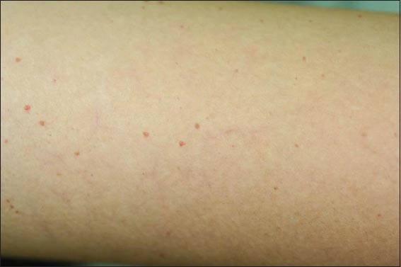


Recently, Miyabe et al. [1] reported two cases of disseminated angiomas on the entire body, which demonstrated several characteristics, e.g. tiny angiomas covering the entire body except for the palms and soles, childhood onset with a gradual increase in number, familial occurrence suggestive of autosomal dominant inheritance, and absence of other systemic disorders such as Fabry’s disease. Histological features in the aforementioned study showed mild hyperkeratosis and vessel ectasia in the papillary dermis. These observations were similar to our own case, although histological evaluation could not be carried out because our patient decisively refused skin biopsy. Familial occurrence was not observed in the present case. Although Miyabe et al. did not exhibit the diagnosis of their cases, there is an old paper termed ‘petechial’ angiomata by Brannen et al. [2], who collected 23 patients that presented multiple small, punctate, vascular lesions on the trunk and extremities. The age of the patients ranged from 20 to 73 years, with a predilection for female. Histological examination showed a simple localized dilatation of a vessel in the subpapillary venous plexus. The cases reported by Miyabe [1] and the present case may be similar to those cases reported by Brannen et al. [2]. Furthermore, linear telangiectasias were scattered on the surface of the extremities and chest. Further accumulation of similar cases is needed and whether those cases may be more common in the Japanese population than was considered should be determined in the future.
CONSENT
The examination of the patient was conducted according to the Declaration of Helsinki principles.
REFERENCES
1. Miyabe C, Okubo Y, Maeda T, Tsuboi R, Mitsuhashi Y, Tiny disseminated angiomas from childhoodEur J Dermatol 2013; 23: 718-9.
2. Brannen M, Nixon RK, Doucette JW, Fosnaugh RP, “Petechial” angiomataArch Dermatol 1961; 83: 386-91.
Notes
Source of Support: Nil
Conflict of Interest: None declared.
Comments are closed.