DOI: 10.7241/ourd.20123.46                                                                                  article in PDF
Our Dermatol Online. 2012; 3(3): 206-209
Date of submission: 18.05.2012 / acceptance: 07.06.2012
Conflicts of interest: None
 
ENIGMATIC NODULES ON THE SKIN – A CASE PRESENTATION

Sunderamoorthy M. Srinivasan

Chettinad Hospital & RI, Chettinad University, Kelambakkam, Tamilnadu, India

Corresponding author: Prof. Sundaramoorthy Srinivasan    e-mail:hamsrini@yahoo.co.in

How to cite an article: Srinivasan S. Enigmatic nodules on skin – a case report. Our Dermatol Online 2012; 3(3): 206-209.


Abstract
Among many skin coloured solitary or multiple nodules of the skin with minimal or no symptoms, cutaneous leiomyomas are unique in its clinical presentation, histopathological features and clinically confusing the dermatologists. Though very rare, few cases of segmental cutaneous leiomyomas have been reported in the medical literature. Cutaneous leiomyomas are rare benign tumors of the skin. Leiomyomas in other regions of the body are on records. Various modalities have been used to relieve pain in such lesions. This presentation highlights the importance of biopsy in all nodular skin lesions of the skin to identify and confirm the disease, plan for the correct treatment, answer questions from the affected patients and to teach dermatology post graduates. Histopathological features of other nodular skin lesions are compared.
 
Key words: leiomyoma; erector pili muscle; „Eel”like nuclei; h-caldesmon; MCUL1


Introduction
Multiple nodular lesions on the skin of human beings create a sense of confusion to the examining dermatologist. He has to rule out many documented nodular skin lesions in an orderly manner before arriving at a final clinically acceptable diagnosis. Even then, he keeps two or more diagnosis for exclusion by the available biochemical and histopathological aids. He will not know till the end what surprise is awaiting him, until when the histopathology shows an entirely different scenario than what he thought of. One among such nodular skin lesions on the human skin, which many dermatologists will never think of while diagnosing clinically is “Cutaneous Leiomyomas”(CL).
 
Case Report
A 30 yrs old married woman, came to the dermatology department with skin colored nodules on her right upper back near the shoulder and over the right forearm (Fig. 1-3). They were slowly evolving, one by one adjoining each other for nearly 3 months. The nodules were skin colored, soft, dome shaped, discrete and occasionally painful. There were seven nodules on the right shoulder and multiple over the right forearm. No signs of inflammation or ulceration were seen around the nodules. The nodules over the forearm were slightly hyper pigmented and also discrete. The skin over the nodules was not pinchable, not attached to the underlying structures. Elsewhere her skin, hairs, nails and mucous membranes were normal. Gynaecological examination was normal.
Clinically the following diagnoses were considered
1. Neurofibroma
2. Neurilemmoma
3. Cutaneous mastocytoma
4. Xanthomas
5. Myxomas
Biochemical studies were normal. Biopsy of the nodules showed the following features [1].
 
1. Bundles of smooth muscle interlacing in the dermis (Fig. 4.5).
2. Muscle bundles were straight.
3. Centrally located, thin, very long, blunt edged, “eel-like” nuclei (Fig. 4,6).
4. Intermingling of varying amounts of collagen.
5. No nuclear hyperchromasia, pleomorphism or mitosis.
The nodules on the right shoulder were excised and the outcome was excellent. After six months there were no signs of any recurrence on the excised site or elsewhere.


Figure 1. Nodules on right shoulder and back
 
Figure 2. Nodules on right shoulder and back (closeup view)
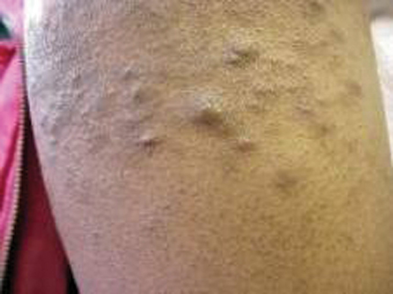
Figure 3. Multiple, slightly hyperpigmented nodules  – Right forearm


Figure 4. Picture of   eel                                                 


  1) Eels are long snake like scaleless marine or
fresh water fishes.
2) They migrate from fresh water to salt water
to spawn.
3) They lack pelvic fins.
 
 
Figure 5. Histopathology of the nodule showing bundles of smooth muscle interlacing in the dermis
Figure 6. Close up view of Figure 4
 
Discussions
Discussion Cutaneous leiomyomas (CL) are benign tumors originating from the erector pilli muscles. They can develop wherever smooth muscle is present. They have equal distribution in both sexes. It has a benign clinical course, and most often presents as multiple cutaneous lesions. Nevertheless, some cases have been described in which single cutaneous leiomyoma appear and even cases in which they appear in families [2-4]. There are reports that associate cutaneous leiomyomatosis to tumors located in other organs, specifically in the uterus and kidneys [5-7]. Pleomorphic adenomas (PA) of the parotid are the most frequently found benign tumors of the major salivary glands [8] and their simultaneous appearance with CL has been reported. First described by Virchow in 1854 [9]. Cutaneous Leiomyomas (CL), are usually not considered while examining any skin coloured nodule with minimal pain, which is sometimes ignored by the patient and missed by the dermatologist. Cutaneous leiomyoma has received little attention in the recent literature. The histopathological features of Cutaneous Leiomyoma is typical and striking to the eyes which are trained to look for it. Hereditary form causes multiple leiomyomas [10], noted by Kloepfer et al in 1958 [11]. Malignant transformation probably does not occur.
Three major types of cutaneous leiomyomas exist:
1. Piloleiomyomas, are believed to arise from the errector pili muscle.
2. Angioleiomyomas, originate from smooth muscle (tunica media) within the walls of arteries and veins.
3. Genital leiomyomas.
 
– They correspond to the histological or anatomic site.
– Menses or pregnancy, temperature and pressure are supposed to be trigger factors for pain [12,13].
– CL are benign tumors that can be exquisitely painful [12,14-16].
– Pathogenesis of pain associated with these lesions is still a mystery.
– The histological findings do not show that prominent nerve fibers are associated with these tumors.
– Others have theorized that specific infiltrating cells may play a role.
– Yet others have suggested that muscle contraction may be pivotal in the induction of pain.
– Genital leiomyomas tend to be the least common of the 3 types.
– Cutaneous leiomyomas with histolopathologic features of uterine symplastic leiomyoma (USL) have also been reported [17,18]. Symplastic leiomyoma is an atypical uterine leiomyoma with cytologic atypia [19].
– Associated morbidity may be due to spontaneous lesional pain, as well as pain evoked by cold and/or tactile hypersensitivity. Additionally, multiple piloleiomyomas have the potential to be cosmetically disfiguring.
– A racial predilection had not been reported.
– The incidence of piloleiomyomas in men and women appear to be equal.
– Symptomatic lesions often necessitate treatment to alleviate discomfort in affected patients.
– Many options are inadequate or create substantial morbidity.
– The search continues for various methods of treatment like CO2 laser ablation, liquid nitrogen cryo, botulinum toxin, nitrous oxide cryo and enucleation [4,20-23].
– A case of cutaneous leiomyomas (CL) arising in a pleomorphic adenoma (PA) of the parotid gland. PA and CL are benign tumours arising from the parotid gland and the erector pilli muscle, respectively [1].
– Cutaneous leiomyomas are more likely to occur in adults than in children.
– Isolated reports of cutaneous leiomyomas in children also exist.
– The most common feature in patients with multiple piloleiomyomas is pain [14,24].
 
Complications:
1. Erythrocytosis associated with skin leiomyomas [8,25].
2. Pain.
 
Distribution patterns:
– Bilaterally symmetric,
– Grouped,
– Dermatomal,
– Linear patterns,
– Piloleiomyomas develop in the superficial dermis, therefore it is fixed to the skin.
 
Recent Research:
– The location of the gene for transmission of dominantly inherited, multiple cutaneous piloleiomyomas associated with uterine leiomyomas in female family members [5].
– As reported by Alam et al., the locus is named MCUL1 (Multiple Cutaneous and Uterine Leiomyomata) [5,26]. Problem in contemporary pathology is:
– The classification and distinction of spindle cell soft tissue tumours of skin. – Markers such as alpha smooth muscle actin (alpha-SMA) and desmin, considered specific for smooth muscle cell (SMC), have been shown to be expressed in variety of fibroblastic and myofibroblastic processes.
 
Conclusion
1. Cutaneous Leiomyomas are rare, most of the time not thought of by dermatologists till the histopathology confirms it. Problem in contemporary pathology is the classification and distinction of spindle cell soft tissue tumors of the skin. 2. Markers such as alpha-smooth muscle actin (alpha-SMA) and desmin, considered specific for smooth muscle cell (SMC), have been shown to be expressed in a variety of fibroblastic and myofibroblastic processes. High-molecular-weight caldesmon (h-caldesmon), one of two isoforms, is reported to be expressed exclusively by SMC and shown to be a specific marker of SMC tumors. 3. h-caldesmon is a specific marker of fully differentiated smooth muscle and that it can serve to differentiate spindled SMC soft tissue tumors of the skin from tumors of myofibroblastic and/or fibroblastic origin.
 
 
Neurofibroma
Neurilemmoma
1. Characteristic round, thin-walled vessels and the mixed nature of the tumor cells.
2. There is no cytologic atypia or mitotic activity.
3. Thin spindle cells associated with thin, wavy collagen bundles.
4. Loosely spaced in clear or mucinous matrix.
1. Small groups of fibrils surrounded by rows of palisaded nuclei.
2. Nuclei in two parallel rows enclosing between them a space nearly homogenous anucleate material.
 Table I.Neurofibroma vs Neurilemmoma (HP showing Verocay body)
 
REFERENCES

1. Holst VA, Junkins-Hopkins JM, Elenitsas R: Cutaneous smooth muscle neoplasms: clinical features, histologic findings and treatment options. J Am Acad Dermatol. 2002; 46: 491-494. 2. Garman ME, Blumberg MA, Ernst R, Raimer SS: Familial leiomyomatosis: a review and discussion of pathogenesis. Dermatology. 2003; 207: 210-213. 3. García Muret MP, Pujol RM, Alomar A, Calaf J, de Moragas JM: Familial leiomyomatosis cutis et uteri (Reed’s syndrome). Arch Dermatol Res. 1988; 280: S29-S32. 4. Sifaki MK, Krueger- Krasagakis S, Koutsopoulos A, Evangelou GI, Tosca AD: Botulinum toxin type A-treatment of a patient with multiple cutaneous piloleomyomas. Dermatology. 2009; 218: 44- 47. 5. Alam NA, Rowan AJ, Wortham NC, Pollard PJ, Mitchell M, Tyrer JP, et al: Genetic and functional analyses of FH mutations in multiple cutaneous and uterine leiomyomatosis, and renal cancer, and fumarate hydratase deficiency. Hum Mol Genet. 2003; 12: 1241-1252. 6. Reed WB, Walker R, Horowitz R: Cutaneous leiomyomata with uterine leiomyomata.- Acta Derm Venerol (Stockh). 1973; 53: 409- 416. 7. Linehan WM, Walther MM, Zbar B: The genetic basis of cancer of the kidney. J Urol. 2003; 170: 2163-2172. 8. Paul M, Attygalle D, Thambirajah M: The origins of leiomyomas. Br J Surg. 1968; 55: 9-14. 9. Virchow R: Ueber Makroglossie and pathologische Neubildung Quergestreifter Muskelfasern. Virchows Arch (Pathol anat). 1854; 7: 126-138. 10. Vellanki LS, Camisa C, Steck WD: Familial leiomyomata. Cutis. 1996; 58: 80-82. 11. Blum P, Jean L: Leiomyome eruptif de Besnier. Bull Soc F Dermatol Syph. 1954; 61: 349-350. 12. Alam M, Rabinowitz AD, Engler DE: Gabapentin treatment of multiple piloleomyoma related pain J Am Acad Dermatol. 2002; 46: S27-S29. 13. Archer CB, Greaves MW: Assessment of treatment for painful cutaneous leiomyomas[letter]. JAm Acad Dermatol. 1987; 17: 141- 142. 14. Batchelor RJ, Lyon CC, Highet AS: Successful treatment of pain in two patients with cutaneous leiomyomata with oral alpha-1 adenoreceptor antagonist, doxazosin. Br J Dermatol. 2004; 150; 775-776. 15. Alam M, Rabinowtz AD, Engler DE: Gabapentin treatment of multiple piloleiomyoma- related pain. J Am Acad Dermatol. 2002: 46(2 Suppl): S27-S29. 16. Thompson JA Jr: Theraphy for painful cutaneousleiomyomas. J Am Acad Dermatol. 1985; 13: 865-867. 17. McGinley KM, Bryant S, Kattine AA, Fitzgibbon JF, Googe PB: Cutaneous leiomyomas lack estrogen and progesterone receptor immunoreactivity. J Cutan Pathol. 1997: 24: 241-245. 18. Suárez-Peñaranda JM, Vieites B, Evgenyeva E, Vázquez-Veiga H, Forteza J: Male genital leiomyomas showing androgen receptor expression. J Cutan Pathol. 2007; 34: 946-949. 19. Archer CB, Whittaker S, Greaves MW: Pharmacological modulation of cold induced pain in cutaneous leiomyomata. Br J Dermatol. 1988; 118: 255-260. 20. Christenson LJ, Smith K, Arpey CJ: Treatment of multiple cutaneous leiomyomas with CO2 laser ablation. Dermatol Surg. 2000; 26: 319-322. 21. Gravvanis A, Kakagia D, Papadopoulos S, Tsoutsos D: Ermal skin template for the management of multiple cutaneous leiomyomas. J Cutan Med Surg. 2009; 13: 1032-1035. 23. Scheinfeld N: The role of gabapentin in treating diseases with cutaneous manifestations and pain. Int J Dermatol. 2003; 42: 491- 495. 24. Abraham Z, Cohen A, Haim S, Greaves MW: Pharmacological Modulation of cold induced pain in cutaneous leioomyomata. Br J Dermatol. 1988; 118: 255-260. 25. Venencie PY, Puissant A, Boffa GA, Sohier J, Duperrat B: Multiple cutaneous leiomyomata and erythrocytosis with demonstration of erythropoietic activity in the cutaneous leiomyomata. Br J Dermatol. 1982: 107: 483-486.

Comments are closed.