Pilar leiomyoma
Niloofar Mehrolhasani
A 30 years old man presented to our dermatology clinic with complaint of multiple firm tender red brown masses on his leg (Figure 1). We did an excisional biopsy. Under high power examination there were interwoven bundles of spindle cells. The nuclei were elongated with blunt ends (cigar shape), so the patient was diagnosed with pilar leiomyoma (Figure 2). We recommended him to use gabapentin for pain relief.
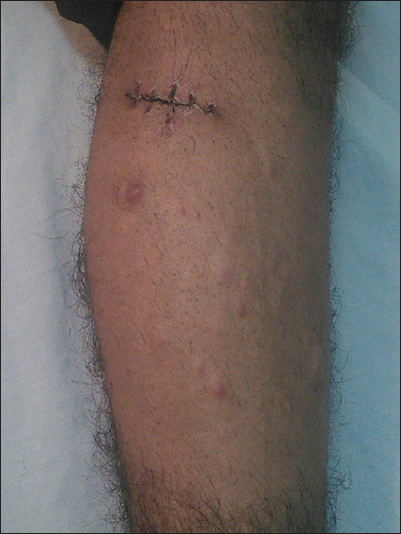
Figure 1:Multiple firm red brown masses on the leg

Figure 2: Interwoven bundles of spindle cells with cigar shape nuclei
The patient’s informed consent was obtained.
Prior to the study, patient gave written consent to the examination and biopsy after having been informed about the procedure.
Pilar leiomyoma is a benign smooth muscle tumor arising from the arrectores pili muscles associated with the hair follicles of the skin [1]. They usually occur as multiple firm dermal nodules located on the extremities and trunk. Leiomyomas usually develop during adolescence or early adult life [2]. Tumours can be painful from compression of cutaneous nerves or because of fibre contraction within the tumour in case of cold weather or emotional stress [3]. The treatment of solitary leiomyoma is surgical excision. In case of multiple leiomyomas, surgery can be done for lesions, which are large and painful. The aim of medical line of treatment is relieving pain. Various drugs have been tried with variable results [4], calcium channel blockers like nifedepine, a-adrenoreceptor blockers (phenoxybenzamine), nitrates, analgesics, antidepressants, and gabapentin. CO2-laser ablation has shown good results [5].
REFERENCES
1. Latoni JD, Neuburg M, Matloub HS, Pilar leiomyoma: a case report and review of the literatureAnn Plast Surg 2000; 45: 662-4.
2. Akay BN, Boyvat A, Heper AO, Unlu E, Congenital pilar leiomyomaJ Am Acad Dermatol 2008; 59: 102-4.
3. Benmously-Mlika R, Ishak F, Ben Jennet S, Hammami H, Badri T, Mokhtar I, Pathologica 2011; 103: 71-2.
4. Raj S, Calonje E, Kraus M, Kavanagh G, Newman PL, Fletcher CD, Cutaneous pilar leiomyoma: clinicopathologic analysis of 53 lesions in 45 patientsAm J Dermatopathol 1997; 19: 2-9.
5. Christenson LJ, Smith K, Arpey CJ, Treatment of multiple cutaneous leiomyomas with CO2 laser ablationDermatol Surg 2000; 26: 319-22.
Notes
Source of Support: Nil
Conflict of Interest: None declared.
Comments are closed.