Erythema multiforme-like impetigo in 10-years-old-girl
Piotr Brzezinski1,2
1Institute of Biology and Environmental Protection, Pomeranian Academy, ul. Arciszewskiego 22A, 76-200 Slupsk, Poland, 2Department of Dermatology, Provincial Specialist Hospital in Slupsk, ul. Mickiewicza 12, 76-270 Ustka, Poland
Corresponding author: Piotr Brzezinski, MD PhD, E-mail: brzezoo77@yahoo.com
Submission: 01.09.2017; Acceptance: 15.10.2017
How to cite this article: Brzezinski P. Erythema multiforme-like impetigo in 10-years-old-girl. Our Dermatol Online. 2017;8(4e):e3.
Impetigo is the most common bacterial skin infection in children two to five years of age. There are two principal types: nonbullous (70% of cases) and bullous (30% of cases). Nonbullous impetigo, or impetigo contagiosa, is caused by Staphylococcus aureus or Streptococcus pyogenes, and is characterized by honey-colored crusts on the face and extremities. Impetigo primarily affects the skin or secondarily infects insect bites, eczema, or herpetic lesions. Bullous impetigo, which is caused exclusively by S. aureus, results in large, flaccid bullae and is more likely to affect intertriginous areas [3].
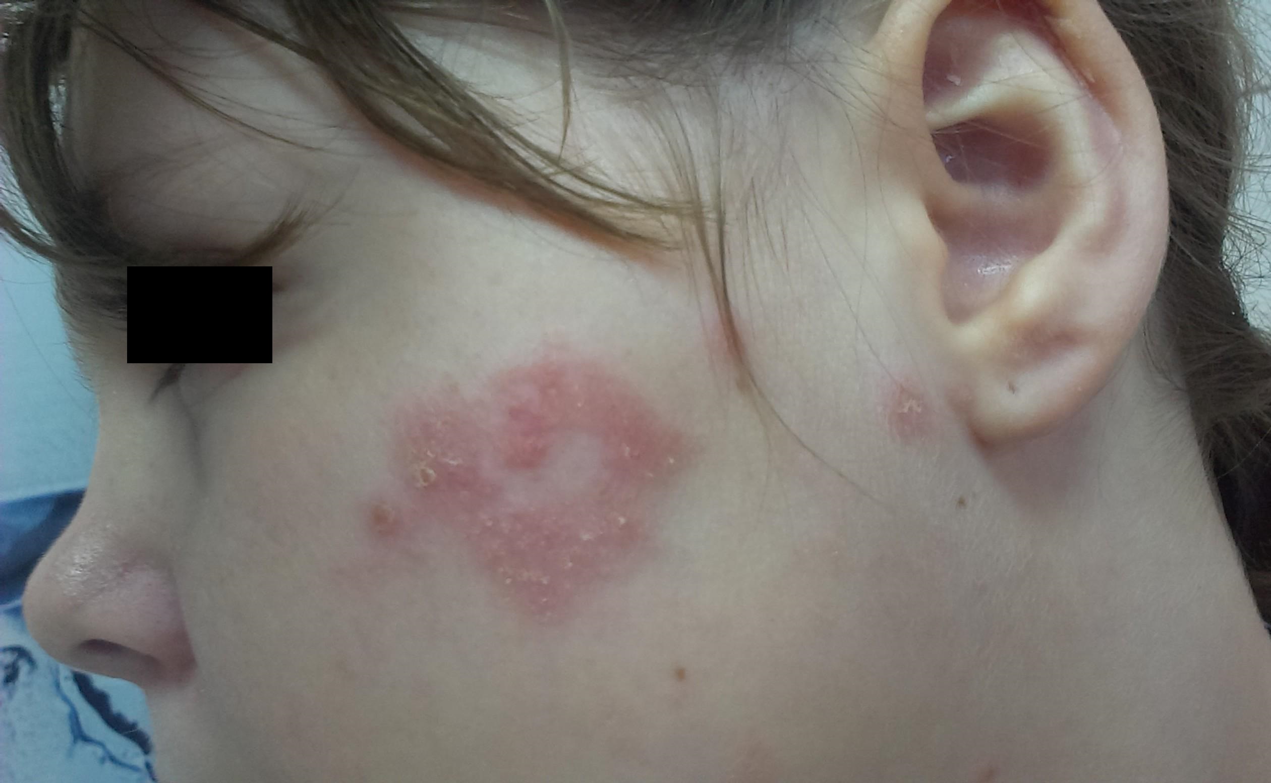
A 10-years-old girl presented with fluid-filled lesions with a small squamous on the face. 5 days duration with history, she has a fever and cough. The lesion resembled impetigo (Fig. 1). In treatment was used Fusidic acid cream.
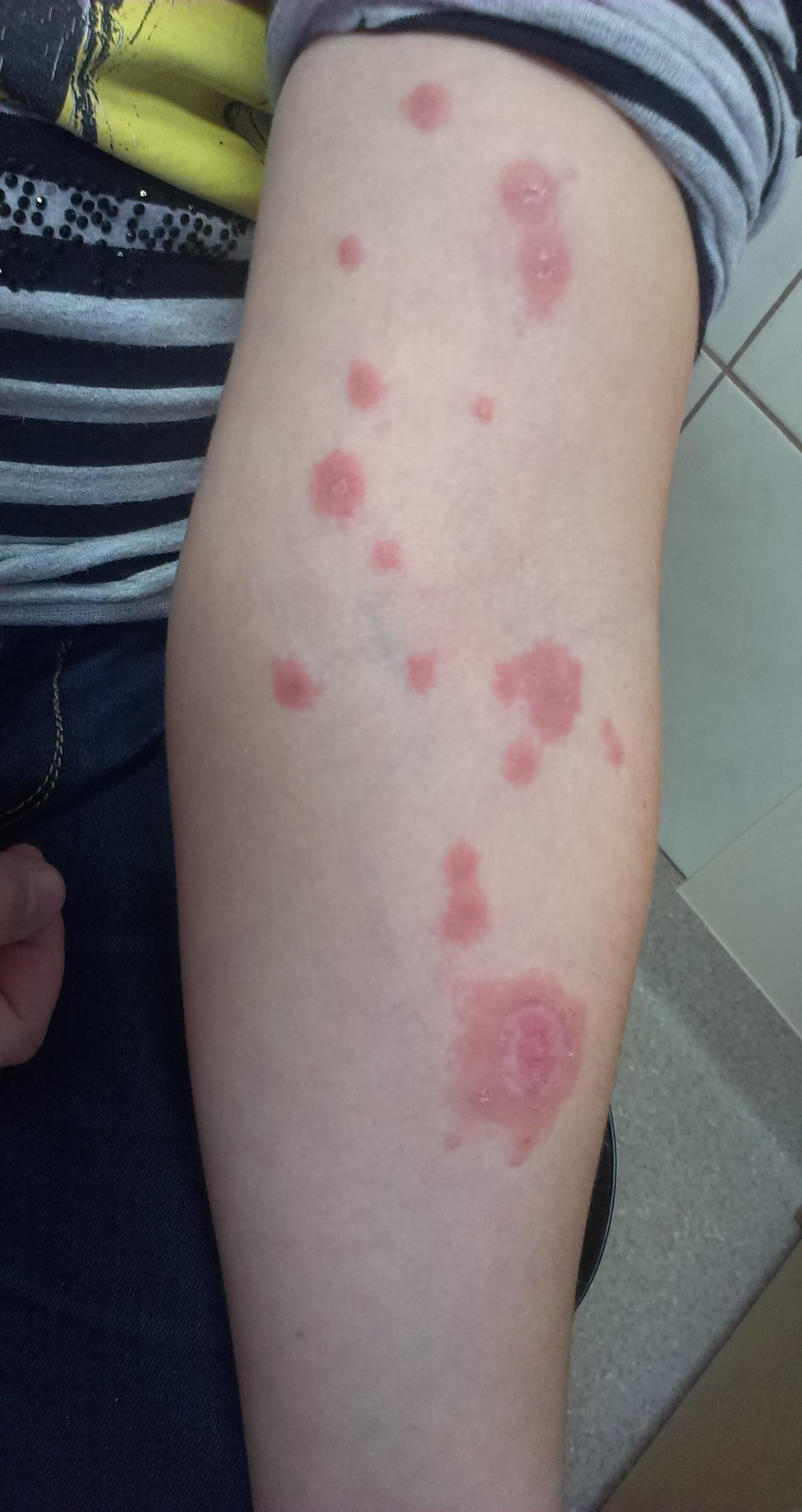
3 days after visiting in the dermatological office a similar lesion occurred on extremities suggesting target lesions of EM (Fig. 2). We know from history that girl had contact with a friend who has had Varicella. The treatment was changed and with successful she responded to topical steroids with antibiotics.
Annular lesions are extremely common in daily clinical dermatological practice, but can be misleading for general practitioners unfamiliar with them [4].
The appearance of annular lesions in childhood is always a diagnostic challenge. The clinical history, appearance and characteristics of lesions by the valuable contribution of histopathology, allow us to know the different entities and separate benign self. The onset injury, lesion characteristics, duration and location, evolution and histological findings to establish a diagnostic orientation and an etiological treatment [4,5].
REFERENCES
1. Bhat YJ, Zeerak S, Baba AN, Hassan I, Wani R. Recurrent erythema multiforme with arthritis – A rare association. Our Dermatol Online. 2017;8:179-82.
2. Gupta M. Hair-dye induced erythema multiforme like allergic contact dermatitis. Our Dermatol Online. 2017;8:177-8.
3. Zitás E, Mészáros J. The most common childhood skin diseases. Our Dermatol Online. 2016;7:213-8.
4. Narváez D, Di Martino Ortiz B, Rodríguez Masi M. Annular skin lesions in childhood: Review of the main differential diagnoses. Our Dermatol Online. 2017;8:75-80.
5. Andres M, Jaworek A, Stramek T, Wojas-Pelc A. Skin reaction to bed bugs bite reflecting erythema multiforme. Case report. Our Dermatol Online. 2015;6:463-5.
Notes
Source of Support: Nil,
Conflict of Interest: None declared.
Comments are closed.