Dark nevus, black lamella and adhesive tape test-a case presentation
Alin Laurentiu Tatu
Faculty of Medicine and Pharmacy, University ,,Dunarea de Jos “, Galati, Romania
Corresponding author: Alin Laurentiu Tatu, MD PhD, E-mail: dralin_tatu@yahoo.com
Submission: 03.03.2017; Acceptance: 10.03.2017
DOI: 10.7241/ourd.2017e.5
How to cite this article: Tatu AL. Dark nevus, black lamella and adhesive tape test-a case presentation. Our Dermatol Online. 2017;8(1e):e6.
A 47 years old man presented with a one month history of changing colour of a nevi after a sun exposure. The lesion was darkened and he was warried about this change. He reported that the lesion is known for about 15 years and was asymptomatic (Fig. 1a). On examination there was a pigmented lesion on the right thigh with a homogenous brown-black pigmentation and no other signs. Dermoscopy revealed a homogenous brown black central pigmentation and a discrete reticular pattern in periphery, some scales on and arround the lesion (Fig. 1b). Due the pigmentation of the rest of the skin the adhesive tape test was proposed: the test involves the repeated sticking of adhesive tape to the lesion, and then tearing it gently off, which results in the removal of the central hyperkeratotic black lamella (Figs. 2a – 2d). After the cornified layer containing pigment(melanin) was removed, the nevi becomed lighter as the surrounded skin and so it was empirically differentiated from melanoma (Figs. 3a and b). A sunscreen with 50 + sun protector factor was recommended, to avoid as possible the sun exposure and also a follow up of the nevi after the sunny season.
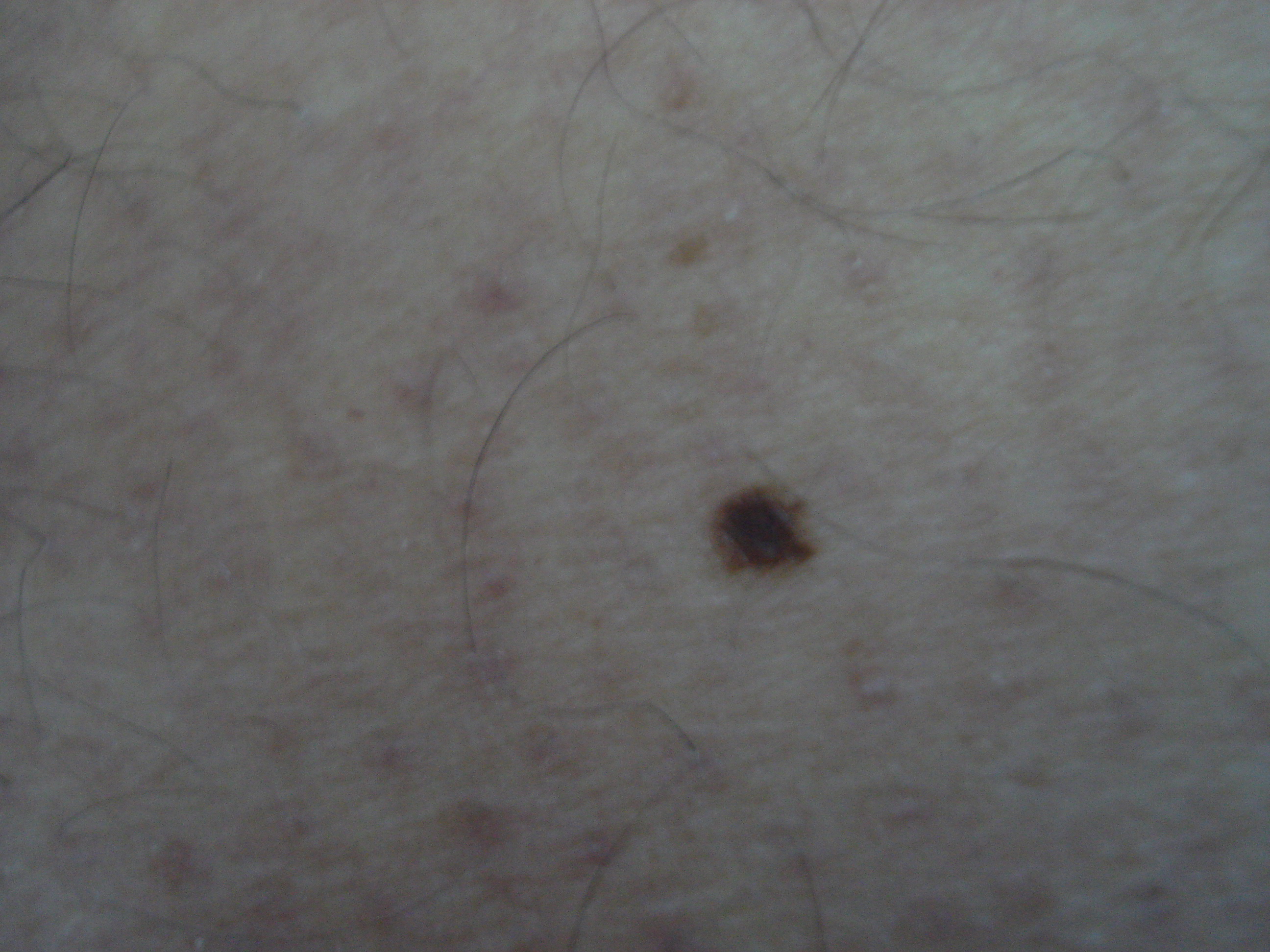
Figure 1a: Dark nevi changed after sun exposure.
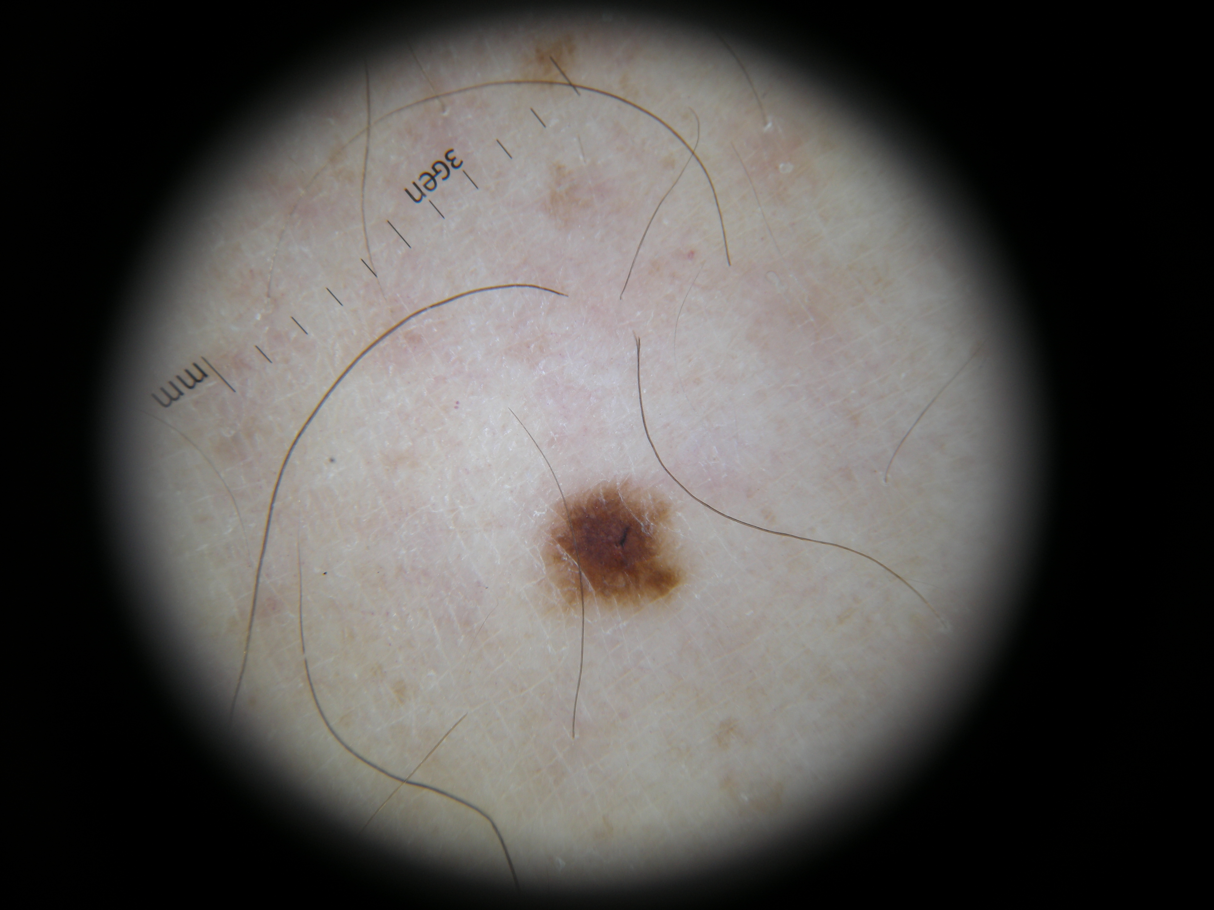
Figure 1b: Brown black homogenous central pigmentation-Dermoscopy.
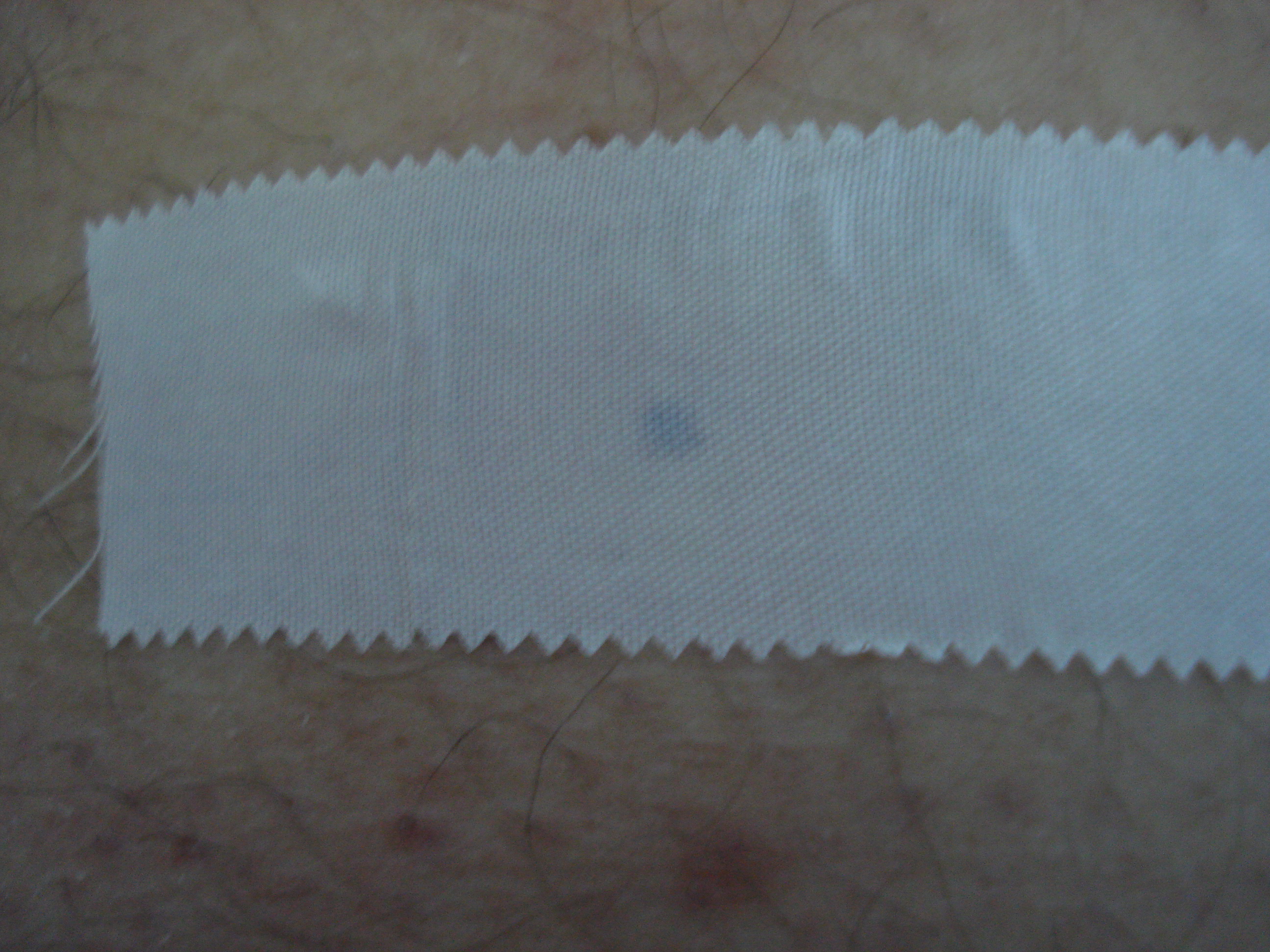
Figure 2a: Adhesive tape test.
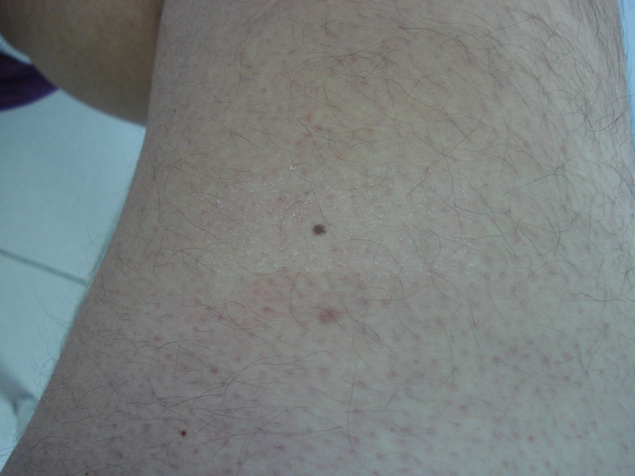
Figure 2b: After gentle tearing the adhesive tape.
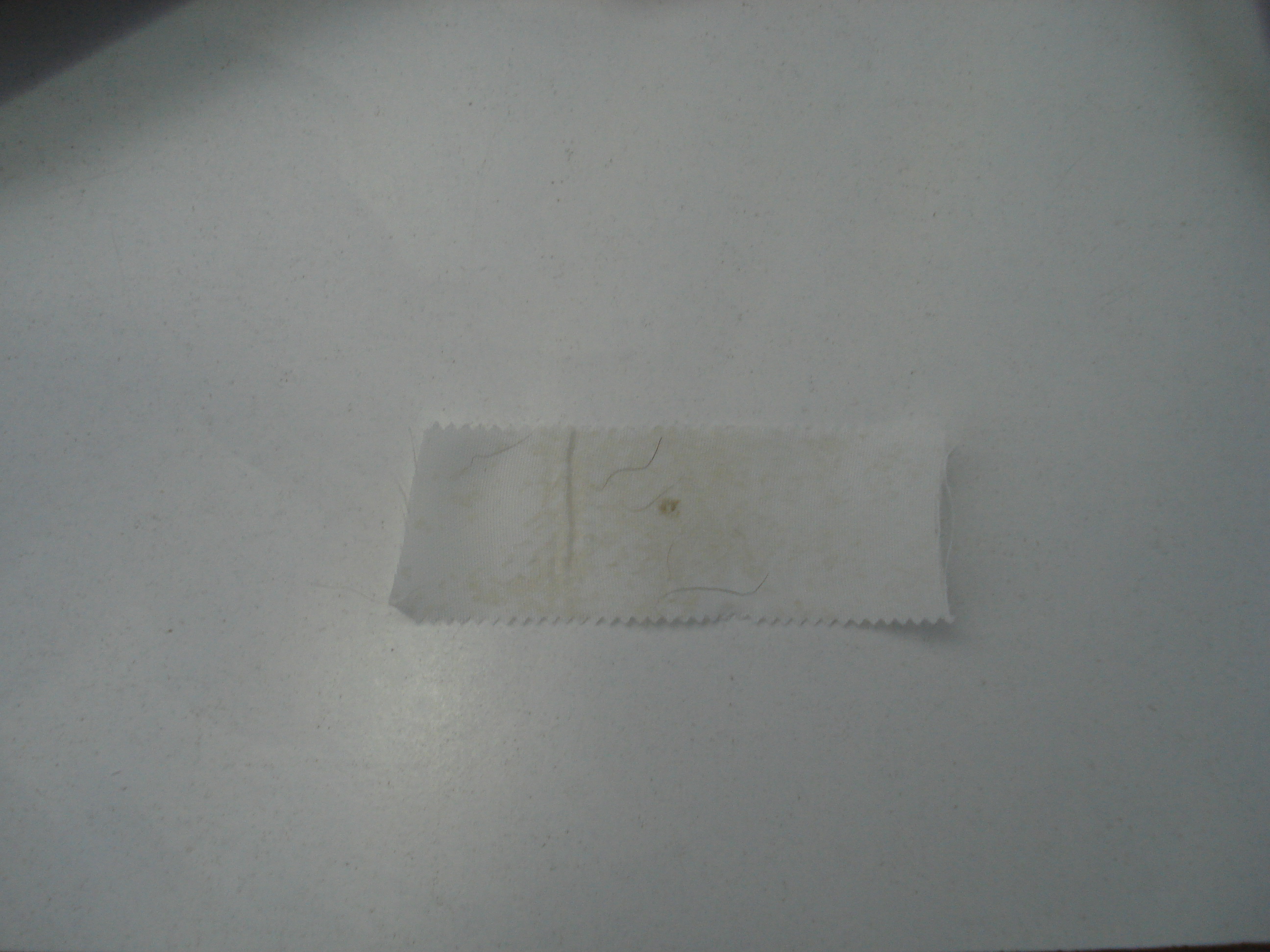
Figure 2c: After tearing on adhesive tape there is a slight pigmentation.
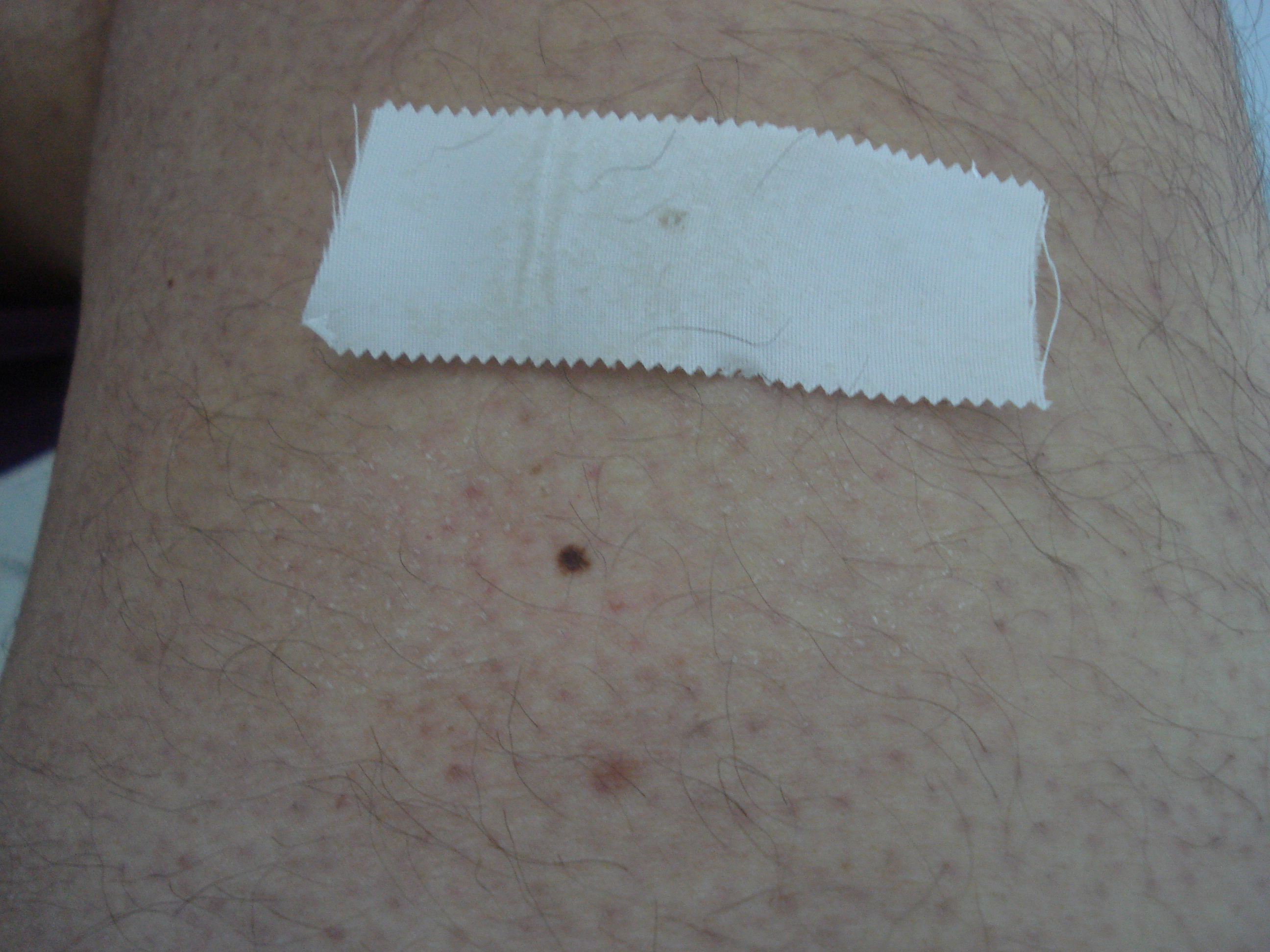
Figure 2d: Adhesive tape test slight depigmentation on nevi and surrounded skin.
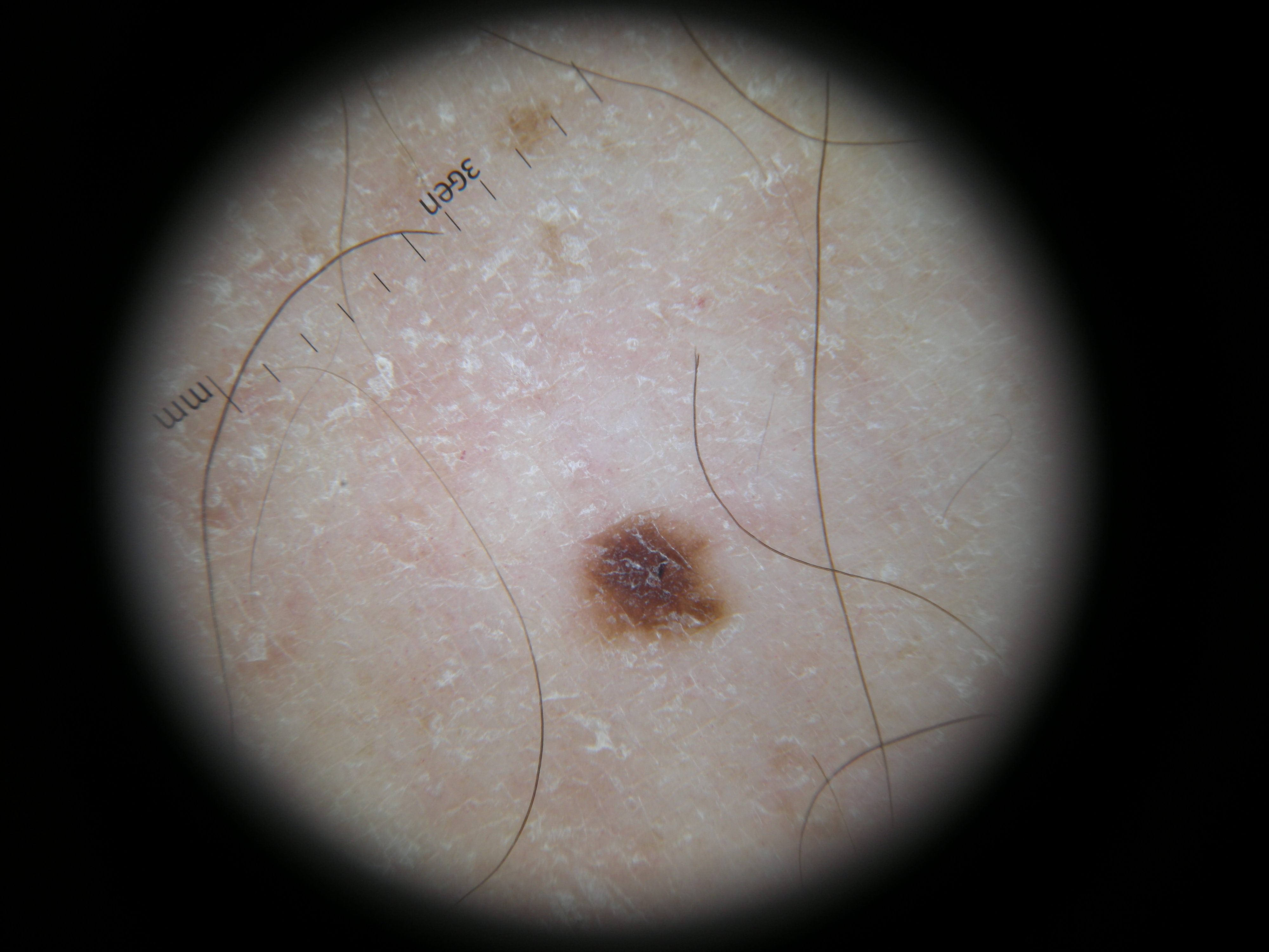
Figure 3a: Dermoscopy after adhesive tape test-a lighter colour on the nevi and scales around
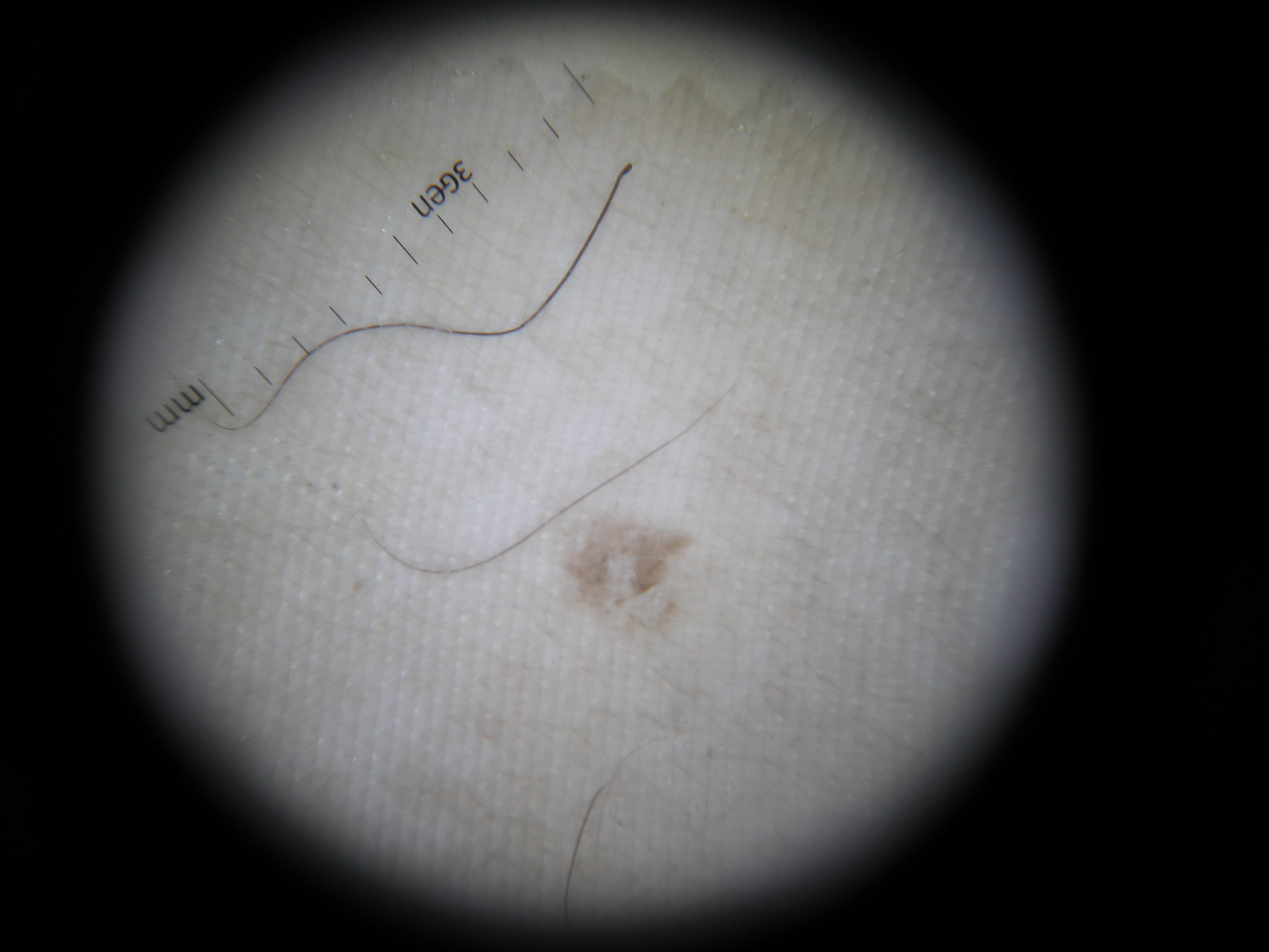
Figure 3b: Central pigment on the tape and slight on periphery-Dermoscopy of the adhesive tape.
Notes
Source of Support: Nil,
Conflict of Interest: None declared.
Comments are closed.