Traumatized blue lesion on the abdominal area
Alin Laurentiu Tatu
Faculty of Medicine and Pharmacy, University ,,Dunarea de Jos “, Galati, Romania
Corresponding author: Alin Laurentiu Tatu, MD PhD, E-mail: dralin_tatu@yahoo.com
Submission: 31.01.2017; Acceptance: 04.02.2017
DOI: 10.7241/ourd.2017e.4
How to cite this article: Tatu AL. Traumatized blue lesion on the abdominal area. Our Dermatol Online. 2017;8(1e):e5.
A 41 years old man was referred pigmented lesion on his inferior abdominal area with a suspicion of a traumatized melanoma (Fig.1) known for about 20 years but slowly growing for about two years. On examination the blue black nodule had dome shaped surface, was firm, mobile with the skin at palpation. There was no bleeding, no local, or regional limphadenopathy, a circular erythema due to a bandage and no other local or general symptoms. At this stage the management options were: surgical excision with safety margins, minimal excision, clinical follow-up or to perform a dermoscopy, because the most common differentials included: nodular melanoma, pigmented traumatied seborrheic keratosis, dermatofibroma, basal cell carcinoma, vascular pigmented lesions like angioma or pyogenic granuloma. Dermoscopy revealed a three millimeters lesion with central white veil and also red and blue circular vascular globules-lacunae in periphery (Fig.2). A presumtive diagnosis of solitary thrombosed limfangioma was made. Due to the possibillity of trauma or bleeding the minimal surgical excision was recommended.
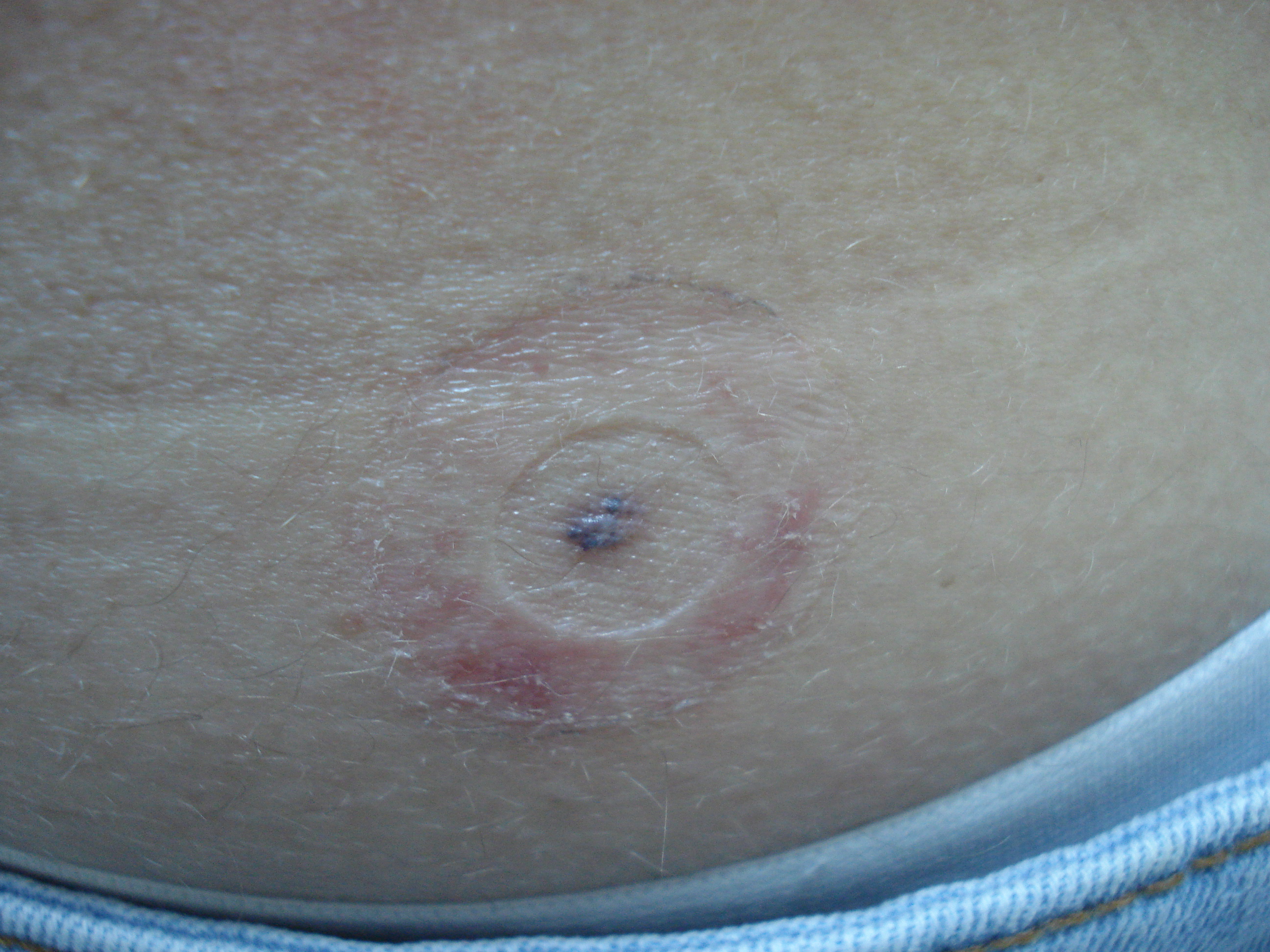
Figure 1: Blue black pigmented abdominal lesion.
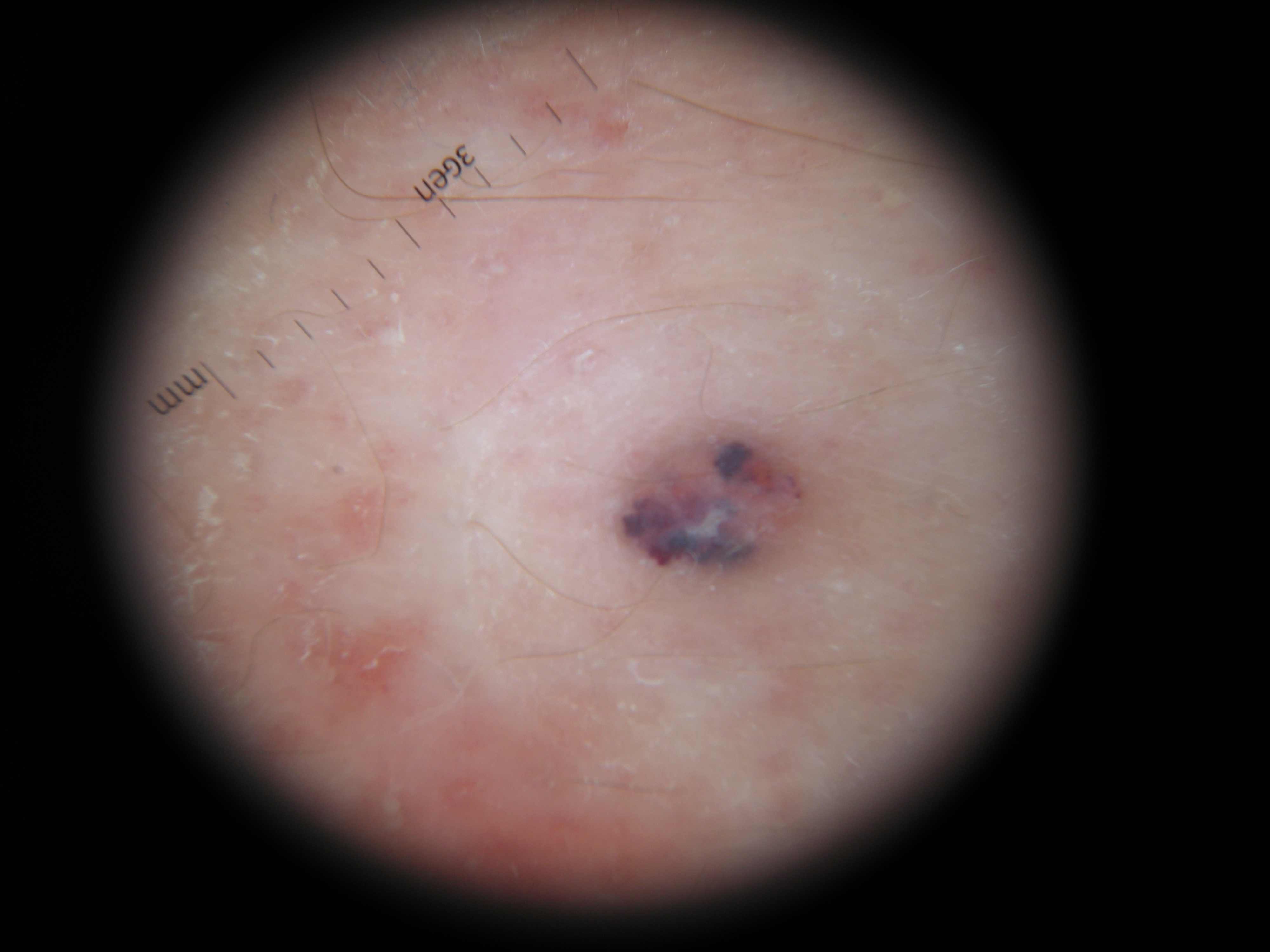
Figure 2: Traumatized limfangioma. Dermoscopy.
Notes
Source of Support: Nil,
Conflict of Interest: None declared.
Comments are closed.