Subungual exostosis associated with an acquired digital fibrokeratoma of the thumb
Maouni Safae , Lakhmiri Myriam, Meziane Mariame, Ismaili Nadia, Senouci Karima
Department of Dermatology-Venereology, Ibn Sina University Hospital, Mohammed V University, Rabat, Morocco
Corresponding author: Dr. Maouni Safae
Submission: 11.12.2019; Acceptance: 17.02.2020
DOI: 10.7241/ourd.2020e.177
Cite this article: Safae M, Myriam L, Mariame M, Nadia I, Karima S. Subungual exostosis associated with an acquired digital fi brokeratoma of the thumb. Our Dermatol Online. 2020;11(e):e177.1-e177.2.
Citation tools:

Copyright information
© Our Dermatology Online 2020. No commercial re-use. See rights and permissions. Published by Our Dermatology Online.
Sir,
Subungual exostosis is a benign osteocartilaginous tumor that occurs in 70 to 80% of cases in the big toe of teenager or young adult. First described by Dupuytren in 1847. Its appearance varies according to its location and age.
A 52 year-old-man, follow-up for neurofibromatosis type 1, presented with a painless periungual nodule of the right thumb, gradually increasing in size over several years (Fig. 1). In dermoscopy, a proximal white area and yellow distal appearance with hyperkeratosis were visible (Fig. 2). X- Ray examination of the right thumb showed a bone outgrowth of distal phalanx (Fig. 3). Surgical removal of the tumour has been performed and histologic examination was consistent with fibrokeratoma, showing an epidermal hyperkeratosis with dermal fibroblasts and collagen bundles.
 |
Figure 1:Periungual nodule of the right thumb. |
 |
Figure 2:Dermoscopy showing a proximal white area and yellow distal appearance with hyperkeratosis ((DermLite DL4, polarized; original magnification: ×10). |
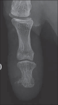 |
Figure 3:X- Ray examination of the right thumb showing the exostosis. |
Cases of subungual exostosis of the fingers are uncommon and described essentially on the thumb of the dominant hand. They are about 1.5 times more in women than in men. Unlike other exostoses, they appear and continue to grow after skeletal maturity (50% of cases in the second or third decade). The etiology is unknown even if a phenomenon of enchondral ossification or peri-ungual soft tissue, secondary to trauma or chronic inflammation is mentioned. Starnes et al. reported several cases involving a balanced translocation t (X; 6).
The association of digital fibrokeratoma and subungual exostosis is unusual, it has been described by Cogrel in 2016 [1]. Both situations may be occur as a periungual nodule and can be favoured by trauma [2], which explains the usual location on the right toenail. In dermoscopy, hyperkeratosis and yellow appearance have been described in both conditions [3], corresponding to epidermal hyperkeratosis in histology, the proximal white area observed in fibrokeratoma represent the dermal fibroblasts with collagen bundles.
The particularity of our observation lies in the association of two benign tumours on a specific genetic field of neurofibromatosis type 1. Dermoscopy as a non-invasive technique can guide the diagnosis in these doubtful cases.
Consent
The examination of the patient was conducted according to the Declaration of Helsinki principles.
The authors certify that they have obtained all appropriate patient consent forms. In the form the patient(s) has/have given his/her/their consent for his/her/their images and other clinical information to be reported in the journal. The patients understand that their names and initials will not be published and due efforts will be made to conceal their identity, but anonymity cannot be guaranteed.
REFERENCES
1. Cogrel O. Excision of a subungual exostosis associated with a fibrokeratoma of the proximal nail matrix of the thumb. Ann Dermatol Venereol. 2016;143:492-3
2. Cohen H, Frank S et al. Subugual exostoses. Arch Dermatol. 1973;107:431-2
3. Salim S, El Meknassi I, Znati K, Meziane M. Acquired ungual fibrokeratoma simulating supernumerary digit:dermoscopic analysis of three cases. J Clin Exp Dermatol. 2019;10:493.
Notes
Source of Support: Nil.
Conflict of Interest: None declared.
Request permissions
If you wish to reuse any or all of this article please use the e-mail (brzezoo77@yahoo.com) to contact with publisher.
| Related Articles | Search Authors in |
|
|
|

Comments are closed.