Porokeratosis: A diagnosis made easy by the dermoscope!
Samia Mrabat , Hanane Baybay, Sabrina Oujdi, Zakia Douhi, Sara Elloudi, Fatima Zahra Mernissi
Department of Dermatology, University Hospital Hassan II, Fes, Morocco
Corresponding author: Samia Mrabat, MD
Submission: Submission: 12.08.2020; Acceptance: 31.10.2020
DOI: 10.7241/ourd.2020e.141
Cite this article: Mrabat S, Baybay H, Oujdi S, Douhi Z, Elloudi S, Mernissi FZ. Porokeratosis: A diagnosis made easy by the dermoscope!. Our Dermatol Online. 2020;11(e):e141.1.
Citation tools:
Copyright information
© Our Dermatology Online 2020. No commercial re-use. See rights and permissions. Published by Our Dermatology Online.
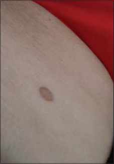
A 56 year-old woman with no medical history presented with an asymptomatic lesion on her right thigh that had been evolving for 20 years. Clinical examination showed an oval shape plaque ; with a slightly erythematous atrophic center surrounded by a hyperkeratotic border (Fig. 1). On dermoscopy, we found a diffuse brown central area of atrophy bounded by irregular double-marginated “white track” border (Fig. 2) consistent with the diagnosis of porokeratosis of Mibelli.
Porokeratoses (PK) are a group of disorders of keratinization characterized by annular lesions surrounded by a characteristic keratotic border which corresponds to a typical histopathologic feature, namely, the coronoid lamella. Though no pathognomonic, the coronoid lamella is the most distinctive feature of the various types of porokeratosis [1]. Dermoscopy is very useful because it reveals specific diagnostic criteria : A peripheral white rim, corresponding to the coronoid lamella, which is the dermoscopic hallmark of porokeratosis [2]. This criteria allows us to confirm the diagnosis without resorting to histology.
CONSENT
The examination of the patient was conducted according to the principles of the Declaration of Helsinki.
REFERENCES
1. Delfi no M, Argenziano G, Nino M. Dermoscopy for the diagnosis of porokeratosis. JEADV. 2004;18:194–5.
2. Moscarella E, Longo C, Zalaudek I, Argenziano G, Piana S, Lallas A. Dermoscopy and confocal microscopy clues in the diagnosis of psoriasis and porokeratosis. J Am Acad Dermatol. 2013;69:e231-3.
Notes
Source of Support: Nil,
Conflict of Interest: None declared.
Request permissions
If you wish to reuse any or all of this article please use the e-mail (brzezoo77@yahoo.com) to contact with publisher.
| Related Articles | Search Authors in |
|
 http://orcid.org/000-0003-3455-3810 http://orcid.org/000-0003-3455-3810 |


Comments are closed.