Fixed pigmented erythema: a potential cause of capricious, atypical and recidivant balanitis
Mohamed El Amraoui , Rachid Frikh, Naoufal Hjira, Mohammed Boui
Department of Dermatology-Venereology, Mohammed V Military Teaching Hospital, Rabat, Morocco
Corresponding author: Dr. Mohamed El Amraoui
Submission: 29.07.2019; Acceptance: 02.10.2019
DOI:10.7241/ourd.2020e.88
Cite this article: El Amraoui M, Frikh R, Hjira N, Boui M. Fixed pigmented erythema: a potential cause of capricious, atypical and recidivant balanitis. Our Dermatol Online. 2020;11(e):e88.1-e88.2.
Citation tools:

Copyright information
© Our Dermatology Online 2020. No commercial re-use. See rights and permissions. Published by Our Dermatology Online.
Sir,
Fixed pigmented erythema (FPE) is a relatively rare toxidermia, elective mucosal involvement is even rarer with difficult diagnosis. At the genital level, the etiological investigation is often oriented towards an STI, especially in young patients. To better illustrate these remarks, we report 3 cases of recurrent and capricious balanitis secondary to FPEs with Sulfonamides and Paracetamol.
Case 1:
60-year-old patient, chronic smoking at a rate of 40 packets/year complicated by chronic obstructive pulmonary disease under inhaled corticosteroids and muco fluidisers. Has consulted for a highly pigmented lesion of the glans, pruriginous, evolving for two years by relapses and remissions (Fig. 1). The STI assessment (syphilitic serology, viral hepatitis B and C and HIV) was negative. The patient admitted that the flares coincided with the outbreaks of his bronchopulmonary disease. The drug investigation has suspected an oral suspension containing Sulfamethoxazole-Trimethoprim with Bromhexine. The diagnosis of an FPE with sulfonamides was retained. Under topical corticosteroids and eviction of sulfonamides, the evolution was favorable with a decline of 2 years.
 |
Figure 1 Fixed pigmented erythema to sulphonamides in the form of a recurrent balanitis. |
Case 2:
Young patient aged 42, with asthma under Salbutamol, consulted for a rash that appeared five days after Sulfamethoxazole-Trimethoprim oral antibiotic therapy for gastroenteritis. Examination revealed erythematous and hyperpigmental balanitis with similar plaques in the left hand (Fig. 2). The diagnosis of EPF with sulfonamides was retained. Under topical corticosteroids and eviction of sulfonamides, the evolution was favorable with a decline of 3 years.
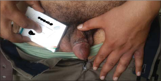 |
Figure 2 Fixed pigmented erythema to sulphonamides with mucocutaneous involvement. |
Case 3:
Young doctor aged 40, without any notable pathological antecedents. Has consulted for an erosive balanitis evolving for two weeks and hindering his sexual intercourse. The interrogation objectified a notion of trauma without notions of risky relationships. The patient was treated with topical antibiotics, antimycotics and topical corticosteroids with a favorable course. One month after the patient presented a recurrence of the symptomatic and dermatological examination showed a centimeter hyperpigmented plaque of the neck. The investigation confirmed the diagnosis of anFPE with Paracetamol.
Toxidermies can only reach the mucous membranes and are real diagnostic puzzles. FPE and less erythema multiforme constitute the two toxidermies that provide exclusive mucosal damage. Some medicines seem to have a particular tropism for certain mucous membranes [1–3]. FPE is an often unrecognized and under diagnosed toxidermy, the antibacterial sulphonamides are a frequent and classic cause, the Elective genital locations are frequent, hence the importance of keeping this diagnosis in mind in front of any recurrent genital dermatosis.
Consent
The examination of the patient was conducted according to the Declaration of Helsinki principles.
The authors certify that they have obtained all appropriate patient consent forms. In the form the patient(s) has/have given his/her/their consent for his/her/their images and other clinical information to be reported in the journal. The patients understand that their names and initials will not be published and due efforts will be made to conceal their identity, but anonymity cannot be guaranteed.
REFERENCES
1. Valeyrie-Allanore L, Lebrun-Vignes B, Bensaid B, Sassolas B, Barbaub A. Fixed pigmented erythema:Epidemiology, physiopathology, clinical features, differential diagnosis and therapeutic management. Ann Dermatol Venereol. 2015;142:701-6.
2. Singhal RR, Sheth NK, Nair PA. Non-pigmented fixed drug eruption caused by ibuprofen. Indian Dermatol Online J. 2019;10:341-3.
3. DiabatéA, Aka RB, Vagamon B, Kourouma HS, GuéI, Kaloga M. Fixed pigmented erythema due to cimetidine. Our Dermatol Online. 2017;8:280-1.
Notes
Source of Support: Nil,
Conflict of Interest: None declared.
Request permissions
If you wish to reuse any or all of this article please use the e-mail (brzezoo77@yahoo.com) to contact with publisher.
| Related Articles | Search Authors in |
|
|
 http://orcid.org/0000-0001-7687-0158 http://orcid.org/0000-0001-7687-0158 |

Comments are closed.