Dermoscopy of Hailey – Hailey disease: Is it useful?
Selma El Kadiri , Hanane Bay Bay, Rhizlane Chaoui, Zakia Douhi, Sara Elloudi, Fatima Zahra Mernissi
Department of Dermatology, CHU Hassan II, Fez, Morocco
Corresponding author: Dr. Selma El Kadiri
Submission: 17.03.2020; Acceptance: 06.04.2020
DOI: 10.7241/ourd.2020e.50
Cite this article: El Kadiri S, Bay Bay H, Chaoui R, Douhi Z, Elloudi S, Mernissi FZ. Dermoscopy of Hailey – Hailey disease: Is it useful? Our Dermatol Online. 2020;11(e):e50.1-e50.2
Citation tools:

Copyright information
© Our Dermatology Online 2020. No commercial re-use. See rights and permissions. Published by Our Dermatology Online.
Sir,
Hailey-Hailey disease or benign familial pemphigus is a rare autosomal dominant blistering dermatosis related to mutations in the ATP2C1 gene [1]. Here we reported a new case with dermoscopic description.
A 45–year-old woman presented a 5-year history of a painful erythematous rash in the skin folds especially in the inframammary area. The patient reported a similar condition to her family members. Physical examination showed irregular, erythematous areas with superimposed thick whitish areas (Fig. 1). The other flexural areas were normal. Dermoscopic examination showed pinkish-white areas in a cloud-like arrangement separated by pink furrows (Fig. 2). Histological examination revealed hyperkeratosis, acanthosis, acantholytic cells giving a dilapidated brick wall appearance with negative immunofluorescence compatible with the diagnosis of Hailey-Hailey disease. The patient was treated with doxycycline 100 mg per day for 3 months showing partial resolution of the symptoms.
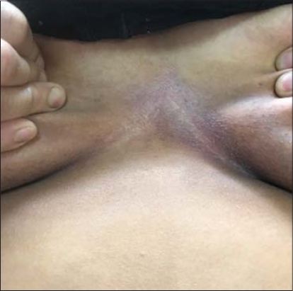 |
Figure 1: Irregular, erythematous areas with superimposed thick whitish areas. |
 |
Figure 2: Dermoscopic image showing pinkish-white areas in a cloud-like arrangement separated by pink furrows. |
The Hailey-Hailey disease is an uncommon genodermatosis characterized by flexural erosions, blisters and warty papules [1]. It can be confused with other flexural disorders such as inverse psoriasis, subcorneal pustular dermatosis, Darier and Dowling Degos diseases [2]. Dermoscopy of Hailey Hailey has been described before and showed a combination of pink and white areas. Irregular whitish areas were separated by pink furrows realizing a cloud pattern. Small erosions and ulcerations were also occasionally visible [3]. In contrary to that, in Darier disease, they are polygonal, star-like, roundish oval, yellowish areas with a whitish peripheral halo [4]. Dermoscopy of inverse psoriasis showed homogeneously distributed “red dots” on an erythematous background with typical bush capillaries [5].
Consent
The examination of the patient was conducted according to the Declaration of Helsinki principles.
The authors certify that they have obtained all appropriate patient consent forms. In the form the patient(s) has/have given his/her/their consent for his/her/their images and other clinical information to be reported in the journal. The patients understand that their names and initials will not be published and due efforts will be made to conceal their identity, but anonymity cannot be guaranteed.
REFERENCES
1. Vasudevan B, Verma R, Badwal S, Neema S, Mitra D, Sethumadhavan T. Hailey-Hailey disease with skin lesions at unusual sites and a good response to acitretin. Indian J Dermatol Venereol Leprol. 2015;81:88–91.
2. Chauhan P, Meena D, Hazarika N. Dermoscopy of Hailey Hailey Disease. Indian Dermatol Online J. 2018;9:139–40.
3. Kelati A. Dermoscopic presentation of Hailey-Hailey disease. J Am Acad Dermatol. 2017;76:S31-3.
4. Lacarrubba F, VerzìAE, Errichetti E, Stinco G, Micali G. Darier disease:Dermoscopy, confocal microscopy, and histologic correlations. J Am Acad Dermatol. 2015;73:e97-9.
5. Micali G. Inverse Psoriasis:From diagnosis to current treatment options. Clin Cosm Investigat Dermatol. 2019;12:953-9.
Notes
Source of Support: Nil.
Conflict of Interest: None declared
Request permissions
If you wish to reuse any or all of this article please use the e-mail (brzezoo77@yahoo.com) to contact with publisher.
| Related Articles | Search Authors in |
|
|
 http://orcid.org/000-0003-3455-3810 http://orcid.org/000-0003-3455-3810 |

Comments are closed.