Axillar trichobacteriosis: Interest of trichoscopy
Selma El Kadiri , Hanane Bay Bay, Rhizlane Chaoui, Zakia Douhi, Sara Elloudi, Fatima Zahra Mernissi
Department of Dermatology and Venerology, University Hospital Hassan II, Fez, Morocco
Corresponding author: Dr. Selma El Kadiri, E-mail: elkadiri-s@hotmail.com
Submission: 18.02.2020; Acceptance: 05.03.2020
DOI: 10.7241/ourd.2020e.29
Cite this article: El Kadiri S, Bay Bay H, Chaoui R, Douhi Z, Elloudi S, Mernissi FZ. Axillar trichobacteriosis: Interest of trichoscopy. Our Dermatol Online. 2020;11(e):e29.1-e29.2
Citation tools:

Copyright information
© Our Dermatology Online 2020. No commercial re-use. See rights and permissions. Published by Our Dermatology Online.
Trichobacteriosis is a frequent superficial bacterial infection of the hair affecting axillae and genital areas and also the scalp [1]. The disease is characterized by yellow nodular concretions, rarely black or red that grow around the hair shaft. Trichomycosis is produced by an aerobic actinomycete called Corynebacterium flavescens or tenuis. This disease characteristic of humid or tropical climates [1]. Here we report a case of a 45n year-old healthy man presented with unpleasant strong axillary odor and dirty bilateral ampits since three months. Physical examination revealed nodular concretions along multiple hair shafts in the axillar area which are firmly adherent (Fig. 1). Trichoscopy revealed yellowish concretions which produced a white-yellow fluorescence under Wood lamp examination (Figs. 2 and 3). The patient was advised to shave the affected areas and administer daily application of topical fusidic acid. Follow-up after six months showed no recurrence of symptoms.
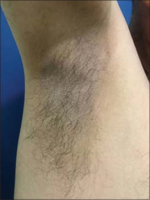 |
Figure 1: Bromohidrosis in a 45 year-old female. |
 |
Figure 2: Yellowish fluorescence under Wood lamp. |
 |
Figure 3: Trichoscopy showing adherent and scattered yellowish concretions. |
Consent
The examination of the patient was conducted according to the Declaration of Helsinki principles.
The authors certify that they have obtained all appropriate patient consent forms. In the form the patient(s) has/have given his/her/their consent for his/her/their images and other clinical information to be reported in the journal. The patients understand that their names and initials will not be published and due efforts will be made to conceal their identity, but anonymity cannot be guaranteed.
REFERENCES
1. Lacarrubba F, VerzìAE, Micali G. Trichoscopy in the differential diagnosis of pseudonits. Skin Appendage Disord. 2019;5:142-5.
Notes
Source of Support: Nil.
Conflict of Interest: None declared.
Request permissions
If you wish to reuse any or all of this article please use the e-mail (brzezoo77@yahoo.com) to contact with publisher.
| Related Articles | Search Authors in |
|
|
 http://orcid.org/000-0003-3455-3810 http://orcid.org/000-0003-3455-3810 |

Comments are closed.